Chapter Summary: Haloalkanes and Haloarenes (CBSE Class XII)
The chapter "Haloalkanes and Haloarenes" is a fundamental topic in organic chemistry for the CBSE Class XII curriculum, introducing students to a crucial class of organic compounds. This chapter typically has a weightage of around 6 marks in the board exams.
Key Concepts
1. Introduction and Classification:
* Haloalkanes: These are organic compounds where a hydrogen atom in an alkane is replaced by a halogen atom. The halogen is attached to an sp^3 hybridized carbon atom.
* Haloarenes: These are aromatic compounds where a hydrogen atom in a benzene ring is replaced by a halogen atom. The halogen is attached to an sp^2 hybridized carbon atom.
* Classification: Compounds are classified as mono-, di-, or poly-halogen derivatives based on the number of halogen atoms. Haloalkanes are further classified as primary (1^\circ), secondary (2^\circ), or tertiary (3^\circ) based on the nature of the carbon atom to which the halogen is bonded.
2. Nomenclature and Nature of the C-X Bond:
* Nomenclature: The chapter covers the IUPAC naming of haloalkanes and haloarenes.
* Nature of C-X Bond: The carbon-halogen bond is polar due to the higher electronegativity of the halogen atom. This makes the carbon atom partially positive (\delta+) and the halogen atom partially negative (\delta-), influencing the reactivity of the molecule. The bond length increases and bond strength decreases as you move down the halogen group (C-F < C-Cl < C-Br < C-I).
3. Preparation of Haloalkanes and Haloarenes:
* Haloalkanes: Common methods include:
* From alcohols, using halogen acids, phosphorus halides, or thionyl chloride.
* From alkanes, via free radical halogenation.
* From alkenes, by addition of hydrogen halides (following Markovnikov's rule or anti-Markovnikov's rule).
* Halogen exchange reactions like Finkelstein and Swarts reactions.
* Haloarenes: Preparation methods include:
* Halogenation of aromatic hydrocarbons in the presence of a Lewis acid.
* Sandmeyer and Gattermann reactions from diazonium salts.
4. Physical Properties:
* Boiling Points: Boiling points of haloalkanes are higher than their parent alkanes due to their polarity and higher molecular mass. The boiling point increases with the size of the halogen and the length of the carbon chain.
* Density: The density of haloalkanes is generally higher than that of water.
* Solubility: Despite their polarity, haloalkanes are only slightly soluble in water because they cannot form hydrogen bonds with water molecules.
5. Chemical Reactions:
* Nucleophilic Substitution Reactions (S_N1 and S_N2): This is a key section of the chapter.
* S_N2 (bimolecular): A single-step reaction where the nucleophile attacks from the side opposite to the leaving group. It is favored by primary (1^\circ) haloalkanes. The rate depends on both the concentration of the substrate and the nucleophile.
* S_N1 (unimolecular): A two-step reaction involving the formation of a carbocation intermediate. It is favored by tertiary (3^\circ) haloalkanes. The rate depends only on the concentration of the substrate.
* Elimination Reactions: Haloalkanes can undergo elimination reactions (dehydrohalogenation) to form alkenes, following Saytzeff's rule (the major product is the more substituted alkene).
* Reaction with Metals: Haloalkanes react with metals like magnesium to form Grignard reagents and with sodium (Wurtz reaction).
* Reactions of Haloarenes: Haloarenes are less reactive towards nucleophilic substitution due to factors like resonance stabilization, partial double bond character of the C-X bond, and the sp^2 hybridized carbon. They undergo electrophilic substitution reactions, where the halogen group is deactivating but ortho- and para-directing.
6. Polyhalogen Compounds:
* The chapter also briefly covers the uses and environmental effects of important polyhalogen compounds like dichloromethane, chloroform, iodoform, freons, and DDT.
Anatomy of Dicotyledonous and Monocotyledonous Plants
ysboiviedonous Stem
lyionocqtyledonous Stem
Vascular System
This chapter introduces the internal structure and functional organisation of higher plants. The study of internal structure of plant is called anatomy.
Meristematic Tissues
Plants have cells as their basic unit, which are organized into tissues, which are then organized into organs. The internal structure of different organs in a plant differs. The monocots and dicots of angiosperms are also anatomically distinct. Internal structures exhibit environmental adaptations as well.
Plant anatomy is primarily concerned with the structure and study of tissue organization. A tissue is defined as “a collection of similar or dissimilar cells that share a common origin and perform a specific function.” Secretory tissue, Permanent tissue, and Meristematic tissues or Meristems are the three main types of tissues. There are two types of tissue in plants: Meristematic tissues or Meristems and Permanent tissue.
Meristematic Tissues
Meristematic tissues contain living cells of various shapes. They have a large nucleus that lacks the vacuole. The cells have no intercellular space between them. These cells can be found in the meristem.
Meristematic tissue cells can actively divide to form specialized structures such as buds of leaves and flowers, tips of roots and shoots, and so on. These cells contribute to the plant’s length and bulkiness.
The term “meristem” was coined by Carl Wilhelm von Nägeli. Meristematic tissue contains undifferentiated cells, which serve as the foundation for specialized plant structures.
Meristematic tissue cells divide actively to form specialized structures such as buds of leaves and flowers, tips of roots and shoots, and so on. These cells contribute to the plant’s overall length and width.
Meristematic tissues are made up of living cells of various shapes. Meristem is the zone where these cells exist.
Plant growth is largely restricted to specialized regions of active cell division known as meristems.
The root apical meristem is located at the tip of the root, whereas the shoot apical meristem is located at the farthest region of the stem axis.
Apical meristems are meristems that form at the tips of roots and shoots and produce primary tissues.
Intercalary meristem is a type of meristem that occurs between mature tissues and is found in grasses and regenerated parts.
Some cells ‘left behind’ from the shoot apical meristem during leaf formation and stem elongation form the axillary bud, which is present in the axils of leaves and forms a branch or a flower.
Both apical and intercalary meristems are primary meristems because they appear early in a plant’s life and help to form the primary plant body.
The secondary or lateral meristem is the meristem that appears later than the primary meristem in the mature regions of roots and shoots of many plants. Fascicular vascular cambium, interfascicular cambium, and cork-cambium are a few examples.
Particular areas of the apical meristem produce vascular tissues, ground tissues, and dermal tissues during the development of the main plant body.
After cell division in both primary and secondary meristems, newly formed cells become specialized and lose their ability to divide; these cells are referred to as permanent or mature cells, and they comprise the permanent tissues.
Classification of Meristematic Tissues
The meristematic tissue can be classified into different types according to its function, position, and origin.
Based on position
Lateral Meristem
It is found on the lateral side of the stems and roots.
It increases the plant’s thickness.
The two lateral meristems are vascular cambium and cork cambium.
These divide either pre-clinically or radially, resulting in an increase in secondary permanent tissues.
Apical meristem
These are found at the tips of the roots and shoots and aid in the growth of the plant’s height.
Cell divisions of various types aid in the growth of cells in the roots and shoots. and promote cellular growth
The apical meristem is divided into two parts:
The promeristem zone is made up of dividing cells (apical initials)
Protoderms (epidermis), procambium (primary vascular tissue), and ground meristem are all found in the meristematic zone (cortex and pith).
Intercalary Meristem
It is found at the intercalary position within the leaves and internodes.
These contribute to the lengthening of the internode.
It is surrounded by grass, monocots, and pines.
It is a component of the apical meristem and contributes to the plant’s peak.
Based on Origin
Primordial meristem
Because the promeristem develops from the embryo, it is also known as the primordial or embryonic meristem. It is found in areas where an organ or a part of the plant body is developing.
Promeristem is a group of initial cells that form the foundation of an organ or plant part. This group is made up of a small number of cells that divide repeatedly to give rise to primary meristem. All other meristems, including the primary meristem, are derived from the promeristem.
Secondary Meristem
Secondary meristems get their name because they develop from permanent cells.
Secondary meristems in the plant body produce secondary tissues and add new cells for effective protection and repair.
A good example of a secondary meristem is phellogen or cork cambium.
Primary Meristem
A promeristem gives rise to a primary meristem, which retains its meristematic activity. It is found at the apex of roots, stems, and leaf primordial, for example, Apical meristem and Intercalary meristem.
Based on Function
Ground Meristem
The cells are quite large and have thick walls.
It contributes to the formation of the cortex, pericycle, and pith.
Meristematic tissue is found in the apices of root systems and shoots and divides continuously.
Protoderm
It is the epidermis, the tissue that covers the outside of the plant.
It protects the plants from mechanical shocks.
Procambium
It is the innermost tissue that gives rise to the xylem and phloem.
It aids in the transport of water and nutrients to various parts of the plant.
Based on the Plane of Division
Mass Meristem
It divides into all planes, forming a mass of cells, such as endosperm.
Rib meristem
It divides in a single plane, resulting in a row of cells. Cortex, for example.
Plate Meristem
It divides into two planes, forming a cell plate. For example, epidermis and epiblema.
Characteristics of Meristematic Tissues
They have a small number of vacuoles.
Meristematic tissue is alive and thin-walled, with the ability to self-renew.
When a cell divides, one cell remains identical to the parent cell, while the others develop specialized structures.
The protoplasm of the cells is very dense, and the meristematic tissues heal an injured plant’s wounds.
Meristematic tissue cells are immature and young.
They do not store food and have a high metabolic rate.
Uses of Meristematic Tissues
They’re alive, and they’re composed of a swarm of rapidly dividing cells.
Tissue is composed of totipotent (all-powerful) cells.
Indefinitely, the cells remain embryonic (immature) and unspecialized.
Vacuoles are typically absent or extremely small if present and cells can be spherical, polygonal, or rectangular in shape.
The nucleus is large and can be found in divisions’ interphase and subphases.
With the exception of mitochondria, other cell organelles are absent or in a non-functional state.
The cell wall is composed of cellulose, which is a homogeneous component, and the cells have a large nucleus and a large number of protoplasms.
Do not keep reserved food materials if orgastic chemicals are missing.
Permanent Tissues
Plants, just like animals are multicellular eukaryotes. This means that they consist of cells, tissues, and organs each with a special unique function to carry out. A cell is the smallest building block of any organism. A group of cells together forms a tissue, and a group of tissue combine to form an organ. Plants belong to the Plantae kingdom, which consists of all organisms which are not plants including fungi and algae.
The plant tissue system is broadly divided into two types: meristematic tissue and permanent tissue(non-meristematic tissue). The meristematic tissue consists of undifferentiated cells(meristematic cells) capable of cell division. These cells can further develop into other tissues and organs that are found in plants. These cells continue to divide till they lose the ability to divide any further and become differentiated. Differentiated plant cells can not divide any further or produce any more cells.
Permanent tissue is defined as a group of living or dead cells formed from the meristematic tissue that has now lost the ability to divide and now have become fixed at a place permanently. These plant tissues take up a specific role after they have lost the ability to divide. The process by which they lose their ability to divide and retain a permanent shape and size and perform a specific function is known as cellular differentiation. They are further divided into two types: Simple Permanent Tissue, Complex Permanent Tissue.
Simple Permanent Tissue
These tissues are also known as homogeneous tissue. They consist of only a single type of cell which has the same origin, structure, and function. They are again classified into three main types:
Parenchyma
The name comes from the Greek words para and enchyma which means beside and tissue respectively. It consists of unspecialized living cells with thin cells. These cells are loosely packed so there are intercellular spaces present in between them. They are generally oval or round in shape. They contain a small number of vacuoles, sometimes it is nonexistential. This is found in all plants and is responsible for the transportation of food and water. The reproductive cells in plants are also Parenchyma.
Characteristics of Parenchyma
They are found in all plants.
The cells in the Parenchyma tissue are living.
The cell walls of Parenchyma tissue are very thin and are made up of Cellulose.
They are generally oval or round in shape.
In the center of the cell, there is a large Vacuole.
There are intercellular spaces present between the cells in this tissue.
These cells have the ability to multiply after they have matured, this property is helpful in the regeneration of damaged plant cells.
Functions of Parenchyma tissue
These tissues are suitable for storage due to the large intercellular spaces in between them. These tissues can store water, fats, oil, etc.
They are capable of transporting nutrition and other chemical products formed in the plant. Some cells are also capable of transporting light through them.
This tissue is responsible for photosynthesis and thus for the general well-being of the plant.
They convert to another type of cell when required for some specific function.
These cells have the ability to multiply after they have matured, this property is helpful in the regeneration of damaged plant cells.
Collenchyma
The name comes from the Greek words colla and enchyma which means gum and tissue respectively. It consists of thin-walled cells. These cells are made up of cellulose and pectin. Due to the presence of pectin, the refractive index of these cells is very high. These cells are very closely packed so there are no intercellular spaces present in this tissue. These tissues are present in the epidermis and vascular bundle of the dicot leaf.
Characteristics
The cells of collenchyma are elongated and spherical or oval in shape.
These cells contain a primary cell wall.
These cells are alive when they mature but not after that.
These cell walls are thick due to the deposition of cellulose and pectin on the cell walls. This thickness is not evenly distributed as the cell wall is thick mostly at the corners but not at the center.
All these cells are arranged very closely so intercellular spaces are not present in this tissue.
The main purpose of these tissues is to provide mechanical support to plants.
Functions
The main purpose of collenchyma is to provide flexibility and support plant growth.
It is mostly present in the growing parts of the plant.
It contains chloroplast and is considered responsible for photosynthesis.
The purpose of collenchyma is to protect leaf margins from tearing.
Sclerenchyma
The name comes from the Greek words Sclerous and enchyma which means hard and tissue respectively. It mostly consists of thick-walled dead cells. They are hard and extremely thick and are uniformly distributed. The main purpose of this is to provide mechanical support. There are no intermolecular spaces present between the tissues. The cell walls become thick due to the deposit of lignin. It is found in shells of nuts, fibers, wood, etc.
Characteristics
It mostly consists of long and narrow cells.
Most of these cells are dead and can not multiply.
The cell wall is thick. They have both primary and secondary cell walls. The secondary cell wall is very thick and has pits that allow the transfer of gases.
The secondary cell walls are made up of cellulose and pectin.
After the cells mature they are mostly dead and they lose the ability to multiply and stretch.
Functions
The main purpose of Sclerenchyma tissue is to provide mechanical support to the plant.
They are present in the protective covering of nuts and seeds.
The xylem vessels and tracheids are also a part of Sclerenchyma tissue.
They form a waxy coating around the leaves to reduce water loss.
Complex Permanent Tissues
Complex Permanent tissues consist of more than one type of cells which have a common origin and which are working together for a common goal as a unit. The main purpose of these complex permanent tissues is the transportation of water and minerals, these tissues are also known as conducting and vascular tissues. It is further divided into two types:
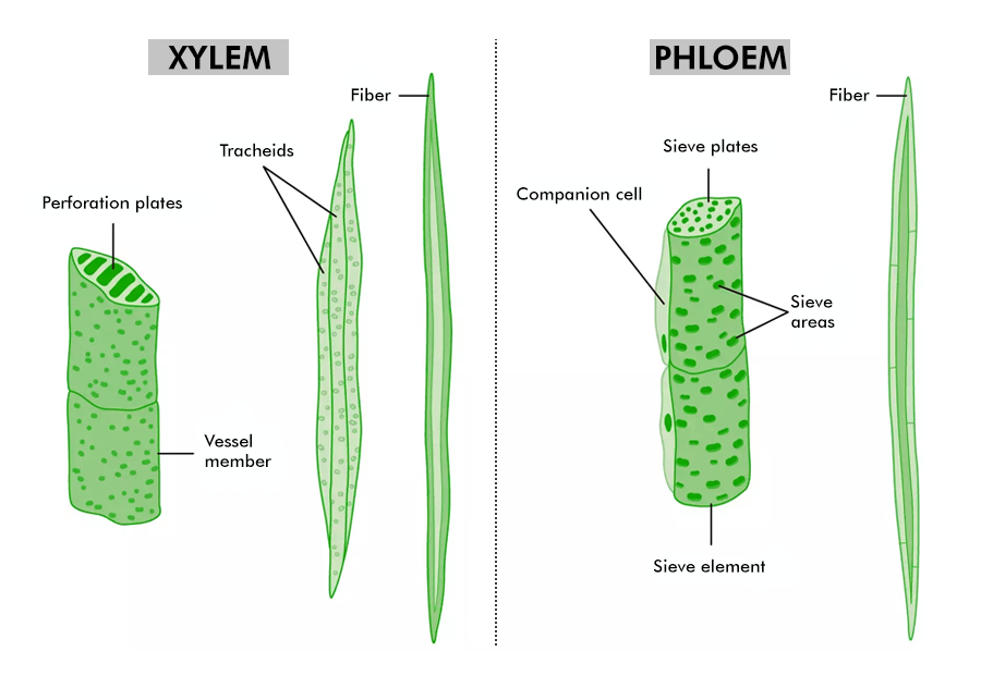
Xylem
The name comes from the Greek word xylos which means wood. It acts as the chief conducting tissue for the plants. It is responsible for the transportation of water and other inorganic solutes. It is divided into tracheids, vessels, xylem fiber, and xylem parenchyma. They are mostly present vertically but they are even present horizontally.
Characteristics
It consists of only living cells.
It has a cell wall, it is very thin and it is made of cellulosic.
These cells have a nucleus and protoplast. These cells are colorless and have large vacuoles.
The nucleus and cytoplasm of xylem tissue migrate into tyloses. Tyloses stores various substances and have a possibility of developing secondary cell walls. The cells which give rise to tyloses are called contact cells.
Functions
The main purpose of xylem tissue is the storage of food materials like starch, fats, and tannins.
They are instrumental in the transportation of water.
When there is a scarcity of water, it helps to reduce damage to the vascular tissues.
They are responsible for repairing and restoring the vessels whenever there is any blockage or formation of air bubbles.
Phloem
The phloem is used to transport food minerals that are dissolved in water. The phloem transports food and minerals both upwards and downwards. It is divided into a sieve tube, Companion cell, Phloem fiber, and Phloem parenchyma.
Characteristics
The cells in the phloem are alive.
It consists of an elongated, tubular structure.
The cell walls are very thin.
Helps in translocation.
It consists of a sieve tube, companion cell, phloem fibers, and phloem parenchyma.
Functions
Carbohydrates produced during photosynthesis are stored in tubes or bulbs which are made of phloem.
The phloem transports sap.
Helps in translocation.
They provide mechanical support to the plants.
Special Permanent Tissues
These tissues are modified structurally and are organized in a particular manner to perform a specific function. For example one of the functions of special permanent tissue is the secretion of some materials like latex. It is further divided into two sub-parts Laticiferous tissues and Glandular tissue.
Glandular Tissue
They are made up of different types of glands. All these lands are formed by single cells or groups of similar cells. The main function of these tissues is to secrete resin, oil, etc. These glands are situated on the epidermis.
Laticiferous Tissues
This is a plant tissue that is responsible for the production and secretion of latex. They are originally formed from meristematic tissue and once they mature they convert to perform specific functions. These tissues consist of thin-walled, elongated cells with multiple nuclei.
Why are Xylem and Phloem called Complex Tissues?
The xylem and phloem are called complex tissues because they consist of different types of cells with specialized functions working together to transport water, nutrients, and sugars throughout the plant. This article will give us a detailed answer to the question “Why are Xylem and Phloem called Complex Tissues?”
Table of Content
Why are Xylem and Phloem Called Complex Tissues?
Merismatic Tissue
Permanent Tissue
Complex Tissue
Characteristics of Xylem and Phloem
Conclusion – Why are Xylem and Phloem called Complex Tissues?
FAQs on Why are Xylem and Phloem called Complex Tissues?
Why are Xylem and Phloem Called Complex Tissues?
Xylem and phloem are termed complex tissues due to their complex structure and specialized functions in plants. The xylem primarily transports water and minerals from roots to leaves, while the phloem distributes sugars produced in the leaves to other parts of the plant.
These tissues comprise various cell types such as vessel elements, tracheids, sieve tube elements, and companion cells, each serving specific roles in nutrient and water transport. The coordinated action of these diverse cell types within the xylem and phloem enables efficient movement of fluids and nutrients throughout the plant, supporting its growth and development.
Also Read: Difference Between Tracheids And Vessels
Types of Plant Tissue
Merismatic Tissue
This is the simple tissue that can continuous cell division. In this type of tissue, growth occurs in the roots and shoot tips of the plant. Based on the region of meristematic tissue in the plant body, we can group the meristems into three types. They are:
Apical Meristems: These meristems are situated at the tips of roots and shoots.
Intercalary Meristems: The intercalary meristems are situated at the internodes or the foundation of the leaves.
Lateral Meristems: These are available on the lateral side of the stem and foundation of a plant.
Permanent Tissue
These tissues are derived from the merismatic tissues and have lost their capacity to divide. This type of tissue can be found in mature plants. They are classified into two types- Simple and complex tissue
Simple Tissue
This is also called homogeneous tissue which consists of a single type of cells. Simple tissues are of three types which are parenchyma, collenchyma, and sclerenchyma.
Parenchyma: These tissues are found in the soft parts of a plant like the roots, stems, and leaves. The cells of this tissue contain huge intercellular spaces between them. Each cell has a vacuole in the middle. The functions of parenchyma tissues are photosynthesis, storage, and assisting the plant with drifting on the water.
Collenchyma: Collenchyma contains oval, round, or polygonal cells with thick walls. The thickness of the walls is due to pectin, cellulose, and hemicellulose. They are intended to offer mechanical help to the plant structure in parts like the petiole of the leaf.
Sclerenchyma: The cells of this tissue are dead. This type of cell lacks a nucleus and cytoplasm. Their cell walls are thickened by lignin and cellulose. Their principal capability is to give mechanical strength to parts of the mature plant.
Complex Tissue
This type of tissue is also known as heterogeneous tissue which consists of different kinds of cells and these differentiated types of cells are coordinated to perform a function. Complex tissues are of two types which are the xylem and phloem.
Complex Tissue
Xylem – Water-Conducting Tissue
The xylem which carries water is made up of tracheary elements like tracheids and vessels mainly. Primary xylem forms during a plant’s early growth from the procambium, while the secondary xylem develops from the vascular cambium later on. It is situated deep within plants and consists of trachea, xylem fibers, tracheids, and xylem parenchyma.
Tracheids and vessels have cylindrical shapes, allowing them to transport water and minerals upwards. Their main job is to move water from roots to stems and leaves, along with nutrients, and provide support to the plant. Xylem tissues are tube-like structures without cross walls and resemble stars. Xylem fibers, smaller in size, are found in the center of vascular bundles and play a role in water replacement lost through transpiration and photosynthesis.
Phloem – Food Conducting Tissue
The phloem which transports food, originates from meristematic cells in the vascular cambium with primary phloem coming from the apical meristem and secondary phloem from the vascular cambium. It’s located in the outer layers of plants, consisting of companion cells, phloem parenchyma, phloem fibers, sieve cells, and sieve tubes. Unlike xylem, phloem moves in both directions and carries food from leaves to other plant parts.
It does not offer structural support. Phloem fibers, larger than those in xylem, are positioned outside vascular bundles, present in stems and leaves, which eventually transport and grow in roots, fruits, and seeds. Phloem main function is to transport transport sugars (primarily sucrose) produced during photosynthesis.
Characteristics of Xylem and Phloem
Phloem and xylem are complex tissues that perform the transportation of food and water to the parts of plants. They are vascular tissues of the plant.
Characteristics
Xylem
Phloem
Structure
Tubular shape with no cross walls which allows a continuous column of water and facilitates rapid transport within the xylem vessels. There are two types, protoxylem and metaxylem depending on the pattern of lignin.
Elongated, tubular shape with thin-walled sieve tubes. The sieve tubes have pores at each end in the cross walls and microtubules that extend between sieve elements allowing the longitudinal flow of material.
Elements
Xylem comprises xylem vessels, fibre and tracheids.
It is located in the centre of the vascular bundle, deep in the plant.
It is located on the outer side of the vascular bundle.
Movement
Movement is unidirectional.
Movement is bidirectional.
Functions
It provides mechanical strength and responsible for transporting water and other nutrients to the plants.
It does not provide mechanical strength and Phloem is living tissue, responsible for transporting food and other organic materials.
Nature of tissue
Xylem consists of dead cells (parenchyma is the only living cells present in the xylem). Xylem constitutes the bulk of the plant body.
Phloem contains living cells (fibres are the only dead cells in the phloem). It forms a small part of the plant body.
Conclusion – Why are Xylem and Phloem called Complex Tissues?
In conclusion, xylem and phloem are termed complex tissues due to their intricate structure and specialized functions in plants. The xylem primarily transports water and minerals from roots to leaves, while the phloem distributes sugars produced in the leaves to other parts of the plant. These tissues consist of various cell types, such as vessel elements, tracheids, sieve tube elements, and companion cells, each playing specific roles in nutrient and water transport. The coordinated action of these diverse cell types within the xylem and phloem enables efficient movement of fluids and nutrients throughout the plant, supporting its growth and development.
Epidermal Tissue System: Its Functions and Tissue in Plant
The epidermal tissue system includes several differentiated cell types including epidermal cells, guard cells, subsidiary cells, and epidermal hairs (trichomes). The epidermis tissue system performs numerous functions: such as preventing water loss, regulating gas exchange, secreting metabolic substances, and absorbing water and mineral nutrients
Tissue System
The body structure of all other developed plants is complex. Similar cells are living together and form a group. It is a group of cells together to perform a specific function, that is, plants have different groups of cells for different forms, which are called tissues.
Sachs (1875) divided the tissues into three systems on the basis of the division of labor in higher-class plants, each system consisting of one tissue or consisting of different tissues. Their function and origin are the same. Tissues are formed by meristems.
Epidermal Tissue System
Ground Tissue System
Vascular Tissue System
Epidermal Tissue System
A group of tissues, no matter how dissimilar they may be, having a similar position and function, is called the epidermal tissue system. The epidermal tissue system is referred to as, the ‘dermal tissue system’. It is the outermost layer of plants. This System consists of the epidermis, stomata, and epidermal outgrowths.
Epidermis
This tissue is made up of epidermal cells and covers all the exposed parts of the plants except the stomatal and stomatal openings. The epidermis is in the form of a continuous layer. The shape and size of the cells are different, due to the cells being adjacent to each other, there are no intercellular spaces. Generally, the epidermis is of one layer (uniseriate), but in many plants, it is multiseriate, such as the Banyan tree. Its cells are made of living parenchyma, in which a large vacuole is found. This vacuole is filled with colorless celli-sap. Chloroplasts are also found in some epidermis. Sometimes these cells have mucilage, and the outer walls are thick due to cutinization and suburbanization. This (cystolith) and silica are also found. the inner and radial of cells. Protects and prevents excess transpiration.
In the leaves of many monocotyledonous plants, some cells of the epidermis are large, thin-walled and these are called bulliform (bubble-like) cells. Many vacuoles are found in these. It is hygroscopic and controls the opening and folding of leaves; as in plants of the Poaceae family.
The guard cells are surrounded by other cells of the epidermis. Their size is often different from other cells of the epidermis. These are called accessory cells or subsidiary cells. The main function of stomata is to exchange gases between the plant and the atmosphere.
The outermost layer of the root is called the epiblema or piliferous layer. Unicellular root hairs emerge from it, which do the work of absorption of water and mineral salts from the soil. Stomata and cuticles are not found in the rhizome.
Stomata
Stomata are found in the aerial parts of leaves and plants. Each stoma is surrounded by two crescent-shaped or kidney-shaped guard cells. The inner wall of guard cells is thick and the outer wall is thin. The guard cells are alive and contain chloroplasts. The guard cells control the opening and closing of the stomata. The guard cells also perform the function of food preparation due to chloroplasts. Due to this, the solution inside it becomes more concentrated, and due to this cells become swollen by absorbing water from the neighboring cells by endosmosis. Due to this, these cells become turgid i.e. due to filling with water, they spread outwards and stomata open. At night the sugar in the cell-sap of these cells is converted into starch. Starch being insoluble in water, the viscosity of the cell membrane in the guard cells is less than in the cells of the neighboring epidermis, thus they lose water by exosmosis and become flaccid. it goes In this way the stomata get closed.
The guard cells are surrounded by other cells of the epidermis. Their size is often different from other cells of the epidermis. These are called accessory cells or subsidiary cells. The main function of the stomata is to exchange gases between the plant and the atmosphere.
Water evaporates through the stomata. Thus the plants get rid of excessive water. And in summer the plants get coolness. The stomata are more numerous in the lower epidermis of the dorsiventral leaves. Their number is relatively less in the upper epidermis. In isobilateral leaves, stomata are equal on both sides. Dorsal leaves are found in dicot plants and isosceles leaves are found in monocot plants. In floating leaves, stomata are found only in the upper epidermis. Like water lilies. Water Stomata are absent in submerged leaves. Like Hydrilla. Sunken stomata are found in desert plants.
The following are the types of leaves based on the presence of stomata:
Astomatic: Stomata are absent in this type of leaf. These are often found in submerged plants, such as Hydrilla and Velisleria, etc.
Hypostomatic: In this type of leaf stomata are mostly found on the lower side of the leaf. For example the leaves of dicotyledons.
Epistomatic: In this type of leaf stomata are found on the upper surface of the leaf. The leaves float on the surface of the water, like a lotus.
Amphistomatic: In this type of leaf almost equal number of stomata are found on both surfaces, like wheat, maize, etc. (all monocotyledonous plants)
Epidermal Appendages
Epidermal outgrowths are growths that appear on the epidermis. There are different varieties depending on the location of the epidermal appendages, which are as follows:
Root hairs
Trichomes
Prickles
Root Hairs
Unicellular outgrowths from the root epidermis or epiblema are known as root hairs. They also help in the absorption of water and minerals from the soil as well as in anchoring.
Trichomes
Many plants develop hairs or trichomes on the epidermis. These may be rigid or soft, unicellular or multicellular branches. These follicles reduce excessive transpiration and protect the plants. Hairs are found on all parts of plants. The role of hair is important in the classification of plants. Following are some of the main types of follicles
Branched: They are found in the form of stars, scales, etc.
Unbranched: In these, all the cells are in a single line; Like a tomato
Nonglandular hair – Non Glandular hair can be unicellular or multicellular, branching or unbranched, stellate or T shaped. These trap air on the surface of the leaves, reducing water loss through transpiration and protecting against extremely hot or cold temperatures.
Glandular hairs are multicellular in nature and release various compounds like mucilage, salt, honey, and so on. Glandular hairs can also function as digestive, aromatic, or stinging glands.
Collectors are glandular trichomes that are either unicellular or multicellular and exude a sticky material. Some trichomes have been adapted to function as bladders for water storage.
Prickles
These are stiffed multicellular epidermal outgrowth. Priclkes are plucked out easily. Prickles help the plant form herbivorous animals. In rose plants prickles are present.
Functions of Epidermal Tissue System
The main function of the epidermis is to protect the inner tissues from injury, heat, cold, and attacks by parasites, fungi, bacteria, etc.
Cuticles, wax, follicles, scales, etc. are found on the epidermis. These prevent excessive evaporation from the plants.
The epidermis acts as a storage of water in desert plants.
Sometimes the epidermis performs the functions of photosynthesis and secretion.
Helps in exchange for gas due to stomata.
Works for absorption of water and mineral salts by root hairs in the root.
Monocot and Dicot Stems – Definition, Structure, Characteristics, Examples
Flowering Plants(Blooming plants) are really characterized into two classes in light of their undeveloped organism, called Monocotyledonous (monocot) plants and Dicotyledonous plants. Allow us to figure out additional about these plants.
We can undoubtedly see the underlying similitudes and varieties in the outer morphology of the bigger living creature, the two plants, and creatures. Essentially, if we somehow happened to concentrate on the inward construction, one to track down a few likenesses as well as contrasts. This part presents you with the inward design and useful association of higher plants. Investigation of the inward design of plants is called life systems. Plants have cells as the fundamental unit, cells are coordinated into tissues and thus the tissues are coordinated into organs. Various organs in a plant show contrast in their inward construction. Inside angiosperms, the monocots and dicots are likewise seen to be physically unique. Inner designs additionally show transformations to different conditions.
Dicotyledonous
The dicotyledons, otherwise called dicots, are one of the two gatherings into which every one of the blooming plants was previously isolated. The name alludes to one of the average qualities of the gathering: specifically, that the seed has two early-stage leaves or cotyledons.
Stems
Stems are normally strong.
Cambium is available.
The quantity of xylem and phloem is two to four and they are recognized by a layer of parenchymatous cells called conjunctive tissue.
Vascular groups in the stem are less organized around and around or ring.
Essence is obvious as is comprised of palisade cells.
Pack sheath missing around vascular groups.
The pericycle is available.
Phloem parenchyma and phloem strands are available.
Monocotyledonous
Monocotyledons regularly alluded to as monocots are endlessly grass-like blossoming plants, the seeds of which normally contain just a single early-stage leaf or cotyledon.
Stems
No cambium thus no auxiliary development in the stem.
The stem is generally empty.
Vascular groups in the stem are dispersed and various.
Phloem parenchyma is missing.
Essence is missing.
Vascular groups are encircled by a sclerenchymatous pack sheath.
The pericycle is missing.
Examples
Examples of Dicotyledonous plants: Tomatoes, Cauliflower, beans, apples, potatoes, and so forth.
Examples of Monocotyledonous (monocot) plants are Maize, Corn, Grass, and Wheat.
Difference between Dicot and Monocot Root
Flowering plants are also called angiosperms. The anatomy of flowering plants includes the organization of cells and tissues within the bodies of flowering plants. Plants are eukaryotic life forms that are portrayed by their capacity to deliver their own food. They give oxygen, food, and medication to other living creatures. The parts of a flowering plant include roots and shoots. These two frameworks are associated with vascular tissue that runs from the root through the shoot. The underground root growth empowers plants to acquire water and supplements from the dirt. The shoot framework permits plants to duplicate and get food through photosynthesis.
The angiosperms are additionally partitioned into monocotyledon and dicotyledon. Monocots vary from dicots in four particular primary highlights: leaves, stems, roots, and flowers. Plants whose seeds contain 1 cotyledon are called Monocots whereas plants whose seeds contain 2 cotyledons are dicots.
Dicotyledonous Root
The internal structure of the dicot root shows the zones mentioned below:
Epidermis
The epidermis has thin-walled, living cells with no intercellular spaces, known as Epiblema. Cells of epiblema protrude out in the form of epidermal root hairs.
Cortex
The cortex has several layers of thin-walled parenchymal cells. The cortex consists of 3 parts:
Exodermis: It is composed of 2 to 3 rows of thick-walled suberized cells. It prevents the exit of water from cortical layers.
General cortex: It is composed of several layers of thin-walled, living, parenchymal cells. It helps in food storage and water conduction.
Endodermis: It is the innermost layer of the cortex. It comprises a single layer of barrel-shaped cells without intercellular spaces.
Radial, as well as tangential walls of endodermal cells, have a deposition of water-impermeable waxy material suberin in the form of Casparian strips.
Endodermis forms a watertight jacket around vascular tissue, so also called an internal biological barrier.
Some endodermis cells near the protoxylem have no Casparian strips, called passage cells or transfusion cells. They allow radial diffusion of water and minerals through the endodermis.
Stele
All tissues on the inner side of the endodermis such as the pericycle, vascular bundle, and pith.
Pericycle
A few layers of thick-walled parenchymatous cells next to endodermis. It shows active cell division. Initiation of lateral roots and vascular cambium during secondary growth takes place in the pericycle.
Vascular Bundles
Radial vascular bundle (Xylem and phloem). Endarch xylem (xylem is on the inner side). Usually, tetrarch means 4 xylem and phloem patches.
Pith
Pith is small or may be completely absent. The parenchymatous cells which lie between the xylem and phloem cells are called conjunctive tissue.
Monocotyledonous Root
The internal structure of the Monocot root shows the zones mentioned below:
Epidermis
The epidermis has thin-walled, living cells with no intercellular spaces, known as Epiblema. Cells of epiblema protrude out in the form of epidermal root hairs.
Cortex
The cortex has several layers of thin-walled parenchyma cells. The cortex consists of 3 parts.
Exodermis: It is composed of 2 to 3 rows of thick-walled suberized cells. It prevents the exit of water from cortical layers.
General cortex: It is composed of several layers of thin-walled, living, parenchyma cells. It helps in food storage and water conduction.
Endodermis: It is the innermost layer of the cortex. It comprises a single layer of barrel-shaped cells without intercellular spaces.
Radial, as well as tangential walls of endodermal cells, have a deposition of water-impermeable waxy material suberin in the form of Casparian strips.
Endodermis forms a watertight jacket around vascular tissue, so also called an internal biological barrier.
Some endodermal cells near the protoxylem have no Casparian strips, called passage cells or transfusion cells. They allow radial diffusion of water and minerals through the endodermis.
Stele
All tissues on the inner side of the endodermis such as the pericycle, vascular bundle, and pith.
Pericycle
It is made from thin-walled parenchymatous cells and is the outermost layer of the stellar system. Monocot roots do not undergo secondary growth. Hence, the pericycle only gives out lateral roots.
Vascular bundles
Radial vascular bundle (Xylem and phloem), Exarch xylem (protoxylem towards periphery metaxylem towards the center). Usually, polyarch means more than 6 xylem and phloem patches.
Pith
It is an enormous, well-developed piece of the monocot root and comprises thin-walled parenchymatous tissue. It contains high measures of starch grains. The parenchymatous cells which lie between xylem and phloem cells are called conjunctive tissue.
Describe the Internal Structure of a Dorsiventral Leaf
Last Updated : 14 Jul, 2022
Flowering plants are also known as angiosperms. The anatomy of flowering plants includes the organization of cells and tissues within the bodies of a flowering plant. Tissues are formed by a group of cells to perform a function. Flowers are known as the reproductive part of the plant since they play the main role in the process of reproduction. Angiosperms are additionally partitioned into monocotyledon and dicotyledon. Monocots vary from dicots in four particular primary highlights: leaves, stems, roots, and flowers. Plants whose seeds contain 1 cotyledon are called Monocots whereas plants whose seeds contain 2 cotyledons are called dicots.
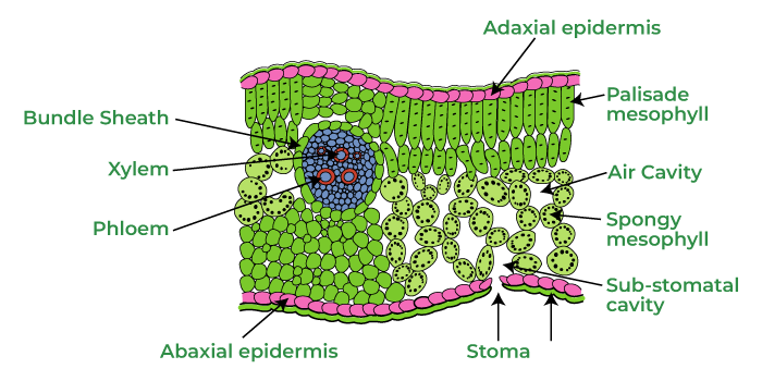
Leaves are important parts of the plant which are mainly involved in photosynthesis. Leaves contain three types of issues which are ground, dermal and vascular. The dermal tissue framework comprises an upper epidermis and lower epidermis. The ground tissue framework that lies between the epidermal layers of the leaf is known as mesophyll tissue. Frequently it is separated into palisade parenchyma on the adaxial (upper) side and spongy parenchyma on the abaxial (lower) side.
A leaf showing this separation in mesophyll is assigned as dorsiventral. It is normal in dicot leaves. The leaves in which mesophyll is not separated like spongy or palisade parenchyma as in monocots, it is called isobilateral. The mesophyll tissue, particularly supple parenchyma cells encase a ton of air spaces. The presence of air spaces is a unique component of springy cells. They work with the vaporous trade between the interior photosynthetic tissue (mesophyll) and the outer air through the stomata.
The vascular tissue framework is made out of vascular packs. The vascular tissue frames the skeleton of the leaf and they are known as veins. The veins supply water and minerals to the photosynthetic tissue.
Anatomy of Dorsiventral (Dicotyledonous leaf)
Dorsiventral leaves can be seen in dicot plants. The internal structure of a dorsiventral leaf shows three particular parts.
Upper epidermis
It is the peripheral layer present on the upper side of the leaf. It is a solitary layer of parenchymatous cells without intercellular spaces. The external walls of the cells are covered with the defensive layer, the fingernail skin. The quantity of stomata present in this layer is less.
Lower epidermis It is the peripheral layer present on the lower side of the leaf. It is a solitary layer of parenchymatous cells without intercellular spaces. The external walls of the cells are covered with cuticles. An impressive number of stomata are available in this layer. Chloroplasts are present just in bean-formed cells, which encompass the stomata openings. With the assistance of stomata, the lower epidermis helps in the trading of gases. Stomata are higher on the abaxial epidermis and the lower epidermis contains stomata without chloroplast.
Mesophyll
Between the upper and lower epidermis, there is a whole mass of ground tissue called mesophyll. It comprises of two various types of parenchyma which are
Palisade parenchyma-This parenchyma is present beneath the upper epidermis. These are extended parenchymatous cells as they have more chloroplasts. In this way, these cells engaged in the course of photosynthesis. The cells have extremely limited intercellular spaces.
Spongy parenchyma-Beneath the palisade parenchyma tissue, springy parenchyma is available. Here, tissues are organized sporadically with intercellular spaces. Thus, cells are for the most part round or oval. This tissue likewise helps in the vaporous trade.
Vascular bundles
These are encircled by thick group sheath cells and should be visible in veins and midrib. These are distinct in size because of reticulate venation. Xylem lies toward the upper epidermis and phloem toward the lower epidermis. The single mid-vein vascular group is bigger, and a few more modest veinlet vascular packs are more modest. More modest vascular packs are uninhibitedly dispersed in mesophyll cells of the leaf.
On account of the isobilateral leaf, the mesophyll tissue in the leaves isn’t separated. It consists of either just springy or palisade parenchyma cells. These sorts of leaves are comparative in an appearance on both sides and, subsequently, are called isobilateral kinds of leaves. On account of the isobilateral leaf, the mesophyll tissue in the leaves isn’t separated. It comprises either just supple or palisade parenchyma cells. These kinds of leaves are comparable in an appearance on both sides and, consequently, are called isobilateral sort of leaves. Most dicots have dorsiventral leaves that are net-veined, including most trees, hedges, garden plants, and wildflowers. Isobilateral leaves. Isobilateral leaves situate themselves lined up with the principal hub and lined up with the bearing of daylight. Isobilateral leaves arrange themselves lined up with the primary pivot and lined up with the bearing of daylight. Most monocots have equal veined isobilateral leaves, including grasses and grass-like plants, lilies, irises, amaryllises, and so forth. Monocot leaf is called isobilateral in light of the fact that both the surfaces of the leaf are similarly green. Leaf, in which the two surfaces are comparable in appearance, are called isobilateral leaves. These are tracked down in monocotyledons. It is because of the presence of light parenchyma.
These leaves situate themselves lined up with the significant hub and lined up with the bearing of daylight. Most monocots have equal veined isobilateral leaves, including grasses and grass-like plants, lilies, irises, amaryllises, and so forth. Epidermis: Monocot leaf has upper and lower epidermis. Isobilateral leaves situate themselves lined up with the fundamental pivot and lined up with the heading of daylight. Most monocots have equal veined isobilateral leaves, including grasses and grass-like plants, lilies, irises, amaryllises, and so on.
Most leaves have specific normal highlights like a covering of an epidermal layer on each surface. The ground tissue that happens between the two epidermal layers is called mesophyll. Vascular groups, normally known as veins, are implanted in the mesophyll. The design and attributes of every one of these layers contrast enormously for dorsiventral and isobilateral leaves.
Diagnostic feature of isobilateral Leaf
Two epidermal layers.
The Cuticle is available on both epidermal layers.
Engine Cells are present in the upper epidermis.
Mesophyll tissue is not separated into palisade and light parenchyma.
Conjoint, insurance, shut-vascular groups.
In an isobilateral leaf, the stomata are available on both the surfaces of the epidermis; and the mesophyll isn’t separated into palisade and light parenchyma. These sorts of leaves are comparable in an appearance on the two sides and, consequently, are called an isobilateral kind of leaves.
On account of the isobilateral leaf, the mesophyll tissue in the leaves isn’t separated. It is composed of either just elastic or palisade parenchyma cells. These sorts of leaves are comparable in an appearance on both sides and, consequently, are called isobilateral kinds of leaves. Dorsiventral passes on situating themselves at a point to the principal pivot and opposite to the heading of daylight. Most dicots have dorsiventral leaves that are net-veined, including most trees, shrubs, garden plants, and wildflowers. Dorsiventral leaves are otherwise called dicot leaves, as they are tracked down in dicotyledons and contain distinct dorsal and ventral sides. It is considered from one another as they contrast from one another in structure as well as appearance. It is separated into palisade parenchyma as well as supple parenchyma.
Anatomy of Isobilateral Leaf
The leaf is the fundamental part of the various pieces of a plant. Leaves carry out two exceptionally fundamental roles, i.e., photosynthesis and happening. Leaf life structures assist us with understanding the tissues and sorts of cells engaged with these cycles and how they work together to perform them effectively. Leaf life structures are read up for significantly two unique kinds of leaves in angiosperms. The dorsiventral leaf and the isobilateral leaf. These two leaves feature the assortment in course of action of tissues and alteration as per the capacities. Dorsiventral leaves show a level arrangement, with all-around separated upper and lower surfaces. The upper surface of the leaf is known as the adaxial or ventral surface. The lower surface of the leaf is known as the abaxial or dorsal surface. An upward cross-part of the leaf displays epidermis (upper and lower), mesophyll, and vascular groups. Isobilateral leaves or monocot leaves are not separated into two particular surfaces. The two surfaces are similarly enlightened by the sun. The leaves show equal venation. The leaf base in many monocots frames a sheath-like construction that covers the stem. The interior association doesn’t show a lot of separation from the dicot leaf.
Key feature of Isobilateral Leaf
On account of the isobilateral leaf, the mesophyll tissue in the leaves isn’t separated. It is composed of either just supple or palisade parenchyma cells. These kinds of leaves are comparative in an appearance on both sides and, subsequently, are called isobilateral sort of leaves. In the dorsiventral leaf, a tissue that is present between the upper and lower epidermis is known as Mesophyll. The chloroplast is present in this tissue and performs the process of photosynthesis. This mesophyll tissue is made up of two types of parenchymatous cells called spongy and palisade parenchyma.
Differences between the isobilateral and dorsiventral Leaf
Dorsi-ventral Leaves
Dorsiventral passes on situating themselves at a point to the principal pivot and opposite to the course of daylight. Most dicots have dorsiventral leaves that are net-veined, including most trees, hedges, garden plants, and wildflowers.
The quantity of stomata is erring on the abaxial epidermis than the adaxial epidermis.
The mesophyll is separated into light and palisade parenchyma.
Vascular groups are enormous and change in size according to the size of veins.
Bulliform cells are missing
Inside, the leaf is separated into the upper and lower epidermis and mesophyll, which are in the middle of between the two epidermises.
The circulation of stomata is normally limited to bringing down the epidermis.
Isobilateral Leaves
Isobilateral leaves situate themselves lined up with the principal pivot and lined up with the course of daylight. Most monocots have equal veined isobilateral leaves, including grasses and grass-like plants, lilies, irises, amaryllises, and so on.
A practically equivalent number of stomata is available on the abaxial and adaxial surfaces.
The mesophyll layer isn’t separated into elastic and palisade parenchyma.
Vascular packs are comparable in size, just the groups close to the mid-vein are enormous.
Bulliform cells are available.
Inside, the leaf is separated into the upper and lower epidermis, and mesophyll, which is in the middle of the two epidermises.
The stomata are distributed on both the epidermises.
Secondary Growth
Term tissue ( French word meaning woven ) was used by N.Grew (1682) the father of plant anatomy. Nageli is regarded as the father of Modern Anatomy. A cell is the functional and structural unit of life. A tissue is a group of cells having a common origin, the same method of development, and function. Anatomy deals with the internal organization of plants. In plants, the terms anatomy and histology have the same meaning. Different organs in plants show differences in their internal structure. Within angiosperms, the monocots and dicots are also seen to be anatomically different. The internal structures show adaptations to diverse environments.
Secondary Growth
Secondary growth is an increase in diameter/girth/circumference due to the addition of secondary tissue(secondary cortex, secondary phloem, and secondary xylem) formed by the activity of vascular cambium in stelar region(region inside pericycle) and cork cambium (phellogen) in extrastelar region (the region outside pericycle i.e., cortex). It is found in the dicot stem, dicot roots, and gymnosperms. Herbs, shrubs, and hydrophytes do not show secondary growth. Monocots rarely show secondary growth(e.g., Aloe, Dracaena, Agave).
Vascular cambium and cork cambium are lateral meristems. Secondary tissues are formed in secondary growth to provide Protection (by cork), mechanical strength (by secondary xylem), conduction of water, and nutrients ( by secondary xylem and secondary phloem). Secondary growth is characterized by an increase in the thickness or girth of the plant. It is caused by cell division in the lateral meristem.
Secondary Growth in dicot Root
It occurs behind root hair zone. In the root, primary cambium is absent. First of all, the cells of conjunctive tissue below phloem region and then, cells of pericycle lying opposite the protoxylem and cells of conjunctive tissue between phloem and xylem regions become meristematic by dedifferentiation and form strips of cambia. Thus, cambium in roots is secondary meristem. The number of such strips depends upon the number of phloem/xylem bundles. These strips extend both ways in between xylem and phloem bundles and finally unite to form a wavy band of vascular cambium. The cambium strip below phloem region becomes active first and divides earlier. Its activity is much faster on the inner side to form secondary xylem. Due to this, the phloem and cambium strips below it are pushed outward making wavy band of cambium a circular one. Now the whole of the cambium becomes active and divides to form secondary xylem. Due to this, the phloem and cambium strips below it are pushed outward making wavy band of cambium a circular one. Now the whole of the cambium becomes active and divides to form 8-10 times more secondary xylem on inner side than secondary phloem on outer side in steler region. It is due to more divisions on the inner side. This causes the pushing of the primary xylem towards the pith and primary phloem towards the periphery. The pith, cortex, and endodermis are fully lost and the primary and old secondary phloem get crushed as the new secondary phloem becomes functional; primary and old secondary xylem persist in old roots. The primary xylem is distinguished by its position. The cells of cambium strips above protoxylem act ad ray initials and divide to form parenchyma on both outer and inner side forming broad, multiseriate parenchymatous primary medullary rays for lateral conduction of water and food. These ray initials are secondary in origin. Secondary medullary rays are more prominent in the root than in the stem. Annual rings are not formed because in the soil, temperature is almost uniform throughout the year.
The secondary tissues formed by the activity of vascular cambium in steler region, exert a pressure on the outer tissue and cause rupturing of epidermis and crushing of cortex and finally peel off. To withstand this pressure, the cells of pericycle become meristematic to form cork cambium(phellogen). It form phellem(cork) on the outside and secondary cortex on the inner side. Cork cambium and secondary cortex together are called periderm which is protective in nature. Lenticels are formed in the cork for aeration. Bark formation is very early in the roots than the stem.
Why Secondary growth is absent in the Monocot root?
In general, monocots do not undergo secondary growth. If they do increase in girth (like palm trees and yucca plants), it does not result in the development of a secondary xylem and phloem, since monocots don’t have vascular cambium. An increase in girth without secondary growth is referred to as anomalous thickening.
Anomalous Secondary Growth
Any deviation from the normal type of secondary growth is called anomalous secondary growth. It is common in plants of tropical regions for example:
Secondary growth in monocots like Dracaena , Agave, Yucca, Aloe by accessory cambia.
Growth in palms is by the activity of primary thickening of apical meristem and persistent leaf bases.
Formation of phloem pockets in the xylem region by the abnormal behaviour of normal cambium in Bignonia, a lianas(woody climber), also called Pyrostegia.
Cambium forms vascular tissues only in the region of vascular bundles.
Significance of Secondary Growth
Secondary growth adds to the girth of the plant. It provides support to increase the weight of the aerial growth.
Secondary growth produces a corky bark around the tree trunk that protects the interior from abrasion, heat, cold and infection.
It adds new conducting tissues for replacing old non-functioning ones as well as for meeting increased demand for long distant transport of sap and organic nutrients.
Cork Cambium
The study of an organism’s internal structure is called anatomy. Histology, or the study of tissue organization and structure, is a component of plant anatomy research. Anatomy reveals the structural adaptation to various settings and reveals the structural differences of various groups of plants.
Tissue is a collection of cells with shared ancestry and typically performing a similar function.
Meristematic tissue is a straightforward tissue made up of clusters of comparable, immature cells that have the ability to divide and create new cells. Apicalmeristems are those that grow at the tips of roots and shoots. In particular, intercalarymeristemsare found between mature tissues in grasses. Both apical and intercalary meristems are primary meristems because they emerge early in a plant’s life and aid in the formation of the main plant body. The term “lateralmeristem” refers to the meristem that grows on the sides of plants and contributes to the growth of their girth. In the primary lateralmeristem, there is intrafascicular cambium. Cork and vascular cambium are secondary meristems.
Permanent tissue refers to cells that have lost the capacity to proliferate and have physically and functionally specialized. Simplepermanenttissues are those with cells that are all similar in structure and function, whereas complicated tissues are those with a variety of cell types. Simple permanent living tissue known as parenchyma is composed of isodiametric cells with thin walls. A substantial central vacuole and nuclei-containing cytoplasm are enclosed within each cell. They can be discovered in the softer, non-woody parts of the stem, root, leaves, fruits, and flowers. They serve as food storage and give plants softer portions turgidity.
Cells in the collenchyma have significantly thicker corners as a result of cellulose, hemicellulose, and pectin. Chlorophyll is frequently oval, spherical, or polygonal in shape. They give the plants’ developing portions, such as young stems, mechanical support.
Cork Cambium
The outer cortical and epidermal layers break down and need to be replaced in order to produce new protective cell layers while the stem continues to girthen as a result of the activity of the vascular cambium. As a result, cork cambium or phellogen, another meristematic tissue, eventually forms, mainly in the cortical area. There are a few levels of phellogen. It is constructed of compact, almost rectangular compartments with thin walls. Cells are severed on both sides by phellogen. Differentiating into cork or phellem are the outer cells. The inner cells develop into a secondary cortex or phelloderm, whereas water cannot penetrate the cork because of suberin deposition in the cell wall. Secondary cortical cells are parenchymatous. The periderm is the aggregate name for phellogen, phellem, and phelloderm.
Pressure accumulates on the remaining peripheral layers as a result of the cork cambium’s activity. These layers eventually phellogen, perish, and peel off. All tissues outside of the circulatory system are referred to as “bark” in a non-technical sense. Cork cambium, which comprises secondary phloem. Periderm and secondary phloem are two of the tissue types that are referred to as bark.
Cork cambium
Structure of Cork Cambium
The periderm’s cork cambium is made up of a single layer of relatively undifferentiated cells. A ring of cells known as the cambium tissue forms on the outside of the plant’s woody tissue and extends the entire length of the mature stem or branch. These undifferentiated cells divide to produce the periderm’s growing cells, particularly the cork cells that make up the branch’s or trunk’s outermost surface.
Early or soft bark is the term used to describe bark that forms early in the season. Late or hard bark forms when the season comes to a close. Give the cell layers of diverse types that make up the bark.
In some areas, the phellogen instead of cork cells removes closely packed parenchymatous cells. Immediately after breaking through the epidermis, parenchymatous cells create lenticels, or apertures resembling lenses. Lenticels allow the transfer of gases that exist between the exterior environment and the stem’s interior tissue. Most woody trees have these.
Functions of Cork Cambium
The cork, a robust protective substance, and secondary cortex are produced by the cork cambium.
In roots and stems, it is in charge of secondary growth that takes the place of the epidermis.
One of the plant’s meristems, a group of tissues made up of embryonic cells from which the plant develops, is the cork cambium. It guards the tree against fungal or bacterial illness.
It stops water from escaping through the bark.
Phellogen (cork cambium) is a meristem that produces periderm tissue.
Difference between Cork Cambium and Vascular Cambium
Cork Cambium
Vascular Cambium
It’s a Meristematic tissue.
It’s also a Meristematic tissue.
It’s a component of tree bark.
It’s present inside the cork cambium.
The cork cambium produces the cork and the secondary cortex.
The vascular cambium produces secondary xylem and secondary phloem.
Cork cambium develops from the secondary lateral meristem.
The vascular cambium develops from the apical meristem.
The stem and root are shielded by the cork cambium, which also stops water loss.
The vascular cambium creates lignified cells and gives the plant its structural support.
Produces the lenticels.
Produces the medullary cells
FAQs on Meristematic Tissues
Question 1: What exactly are meristematic tissues?
Answer:
Meristematic tissue is plant tissue that can divide actively throughout its life. Nageli coined the term meristem (1858). Meristems are found in plant apex, root, leaf primordia, vascular cambium, cork cambium, and so on.
Question 2: What are apical meristems?
Answer:
These are found in the tips of roots and shoot at opposite ends of the plant axis. Cell division and subsequent cellular enlargement in these areas extend the plants above and below ground parts. Meristems also influence the shapes of mature plants because they lay down the patterns for subsequent growth.
Question 3: What exactly is a primary meristem?
Answer:
The cells produced by apical meristem divisions quickly differentiate into three zones of distinct tissues that differentiate below the apical meristems. The protoderm, procambium, and ground meristem are the primary meristems, also known as transitional meristems. They give rise to the primary plant body’s tissue systems.
Question 4: What are some of the characteristics of meristematic tissues?
Answer:
Because when a cell divides, one cell remains identical to the parent cell while the others divide to produce specialized structures, it has the ability to self-renew.
They have the fewest number of vacuoles.
Meristematic tissues are thin-walled, living tissues.
Question 5: What is the Apical Cell Theory?
Answer:
Nageli proposed this theory (1858). The shoot apical meristem, according to this theory, is made up of a single apical cell. This theory applies to higher algae, bryophytes, and many pteridophytes but not to higher plants (i.e., gymnosperms and angiosperms).
FAQs on Permanent Tissue
Question 1: What are the Special Permanent tissues?
Answer:
These tissues are modified structurally and are organized in a particular manner to perform a specific function. For example one of the function of special permanent tissue is secretion of some materials like latex. They are originally formed from meristematic tissue and once they mature they convert to perform specific function. These tissues consist of thin-walled, elongated cells with multiple nuclei.
Question 2: How is the Plant tissue system divided?
Answer:
A cell is the smallest building block of any organism. A group of cells together forms a tissue, and a group of tissue combine to form an organ. The plant tissue system is broadly divided into two types: meristematic tissue and permanent tissue(non-meristematic tissue).
Question 3: Define Meristematic tissue.
Answer:
The meristematic tissue consists of undifferentiated cells (meristematic cells) capable of cell division. These cells can further develop into other tissues and organs that are found in plants. These cells continue to divide till they lose the ability to divide any further and become differentiated.
Question 4: Define Permanent tissue.
Answer:
Permanent tissue is defined as a group of living or dead cells formed from the meristematic tissue that has now lost the ability to divide and now have become fixed at a place permanently. These plant tissues take up a specific role after they have lost the ability to divide. The process by which they lose their ability to divide and retain a permanent shape and size and perform a specific function is known as cellular differentiation.
Question 5: Explain the classification of complex permanent tissues.
Answer:
The main purpose of these complex permanent tissues is the transportation of water and minerals, due to these tissues are also known as conducting and vascular tissues. It is further divided into two types:
Xylem: It acts as the chief conducting tissue for the plants. It is responsible for the transportation of water and other inorganic solutes. It is divided into tracheids, vessels, xylem fiber and xylem parenchyma.
Phloem: The phloem is used to transport food minerals that are dissolved in water. It transports food and minerals both upwards and downwards. It is divided into a sieve tube, Companion cell, Phloem fiber, and Phloem parenchyma.
FAQs on Why are Xylem and Phloem called Complex Tissues?
Why are Xylem and Phylum called Conducting Tissues ?
Xylem and phloem are called conducting tissues because they transport water, nutrients, and food throughout the plant.
Why is Xylem Called Dead Complex Tissue?
Xylem is called a dead complex tissue because its main components, tracheids and vessels, are non-living at maturity.
What are the Constituents of Phloem?
The constituents of phloem include companion cells, phloem parenchyma, phloem fibers, sieve cells, and sieve tubes.
Why is Xylem Known as Water-Conducting Tissue?
Xylem is called water conduction tissue since it transports water molecules from the roots to the parts of the plants.
Why is Phloem Known as Food Conducting Tissue?
Phloem is called food conducting tissue since it transports food and nutrients from leaves to other parts of the plant.
What are the Characteristics of Xylem and Phloem?
Xylem is known for its tubular structures and role in transporting water and minerals upward, while phloem is recognized for its sieve tubes and function in carrying food throughout the plant.
What are the Functions of Xylem and Phloem?
The main function of xylem is to transport water and minerals from roots to stems and leaves, while phloem’s primary role is to transport food produced in leaves to other parts of the plant.
FAQs on Epidermal Tissue system
Q: What is the most important function of trichomes?
Answer:
The epidermal cells have hair-like structures referred to as trichomes. These hairs or trichomes support in protecting the plant from sunlight and outer injuries.
Q: What are the stomata?
Answer:
Stomata are cell structures in the epidermis of tree leaves and needles that help plants exchange carbon dioxide and water with the atmosphere.
Q: What is the function of the epidermal tissue system in plants?
Answer:
The main function of the epidermis is to protect the inner tissues from injury, heat, cold, and attacks by parasites, fungi, bacteria, etc.
Q: Describe guard cells?
Answer:
Guard cells are kidney-shaped cells that surround the stomata. They regulate the opening and closing of the stomata.
FAQs
Question 1: What are the differences between Monocot and Dicot roots?
Answer:
Features
Monocot roots
Dicot roots
Xylem
Polyarch
Usually tetrarch
Pith
Large at the center
Pith is usually absent
Metaxylem
Metaxylem vessels are generally circular in cross section
Metaxylem vessels are generally polygonal in cross section
Conjunctive tissue
It is sclerenchymatous in maize
It is usually parenchymatous
Secondary growth
There is no secondary growth
Secondary growth is present
Shape of xylem
Round or oval shape.
Angular or polygonal shape.
Vascular tissues
More number of xylem and phloem
Less number of xylem and phloem
Cortex area
Wide cortex area
Narrow cortex area
Example
Banana, Palm
Pea, beans
Question 2: What are the functions of roots?
Answer:
Roots carry out different roles that are important for the endurance of the plants. They are a fundamental or coordinated framework that helps the plant in:
They support the plant body, guaranteeing that it stands erect.
The primary capability of the roots is to absorb water and break up minerals from soil which helps during photosynthesis.
Plants store food as starch in the leaves, shoots, and roots. Examples; carrots, radish, beetroot, and so on.
They play important role in the method of reproduction. For example, new plants emerge from crawling even stems called sprinters (stolons) in jasmine, grass, and so on. This sort of proliferation is called the vegetative spread.
Biological Function: They actually take a look at soil disintegration and give food, and furthermore territory to different organic entities.
Question 3: Why does there no secondary growth occur in monocot roots?
Answer:
Expansion in thickness of stem because of the development of lateral tissues is called Secondary growth. It happens generally in dicotyledons. It happens by the arrangement of horizontal meristems like vascular cambium and cork cambium. Vascular cambium is liable for the intra-stelar auxiliary development and cork cambium is responsible for extra stelar optional development. Secondary growth does not occur in monocot plants as they lack cambium in the vascular bundle between xylem and phloem.
Question 4: Which tissue is present more often in monocot roots than in dicot roots?
Answer:
Conjunctive tissues are masses of parenchymatous or sclerenchymatous cells that are available between the xylem and phloem packs in the vascular tissue. The amount of conjunctive tissue is more in monocot roots due to the presence of a large number of vascular bundles when compared to dicot roots. In dicot plants, the conjunctive tissues along with the pericycle lead to the vascular cambium during auxiliary development. No cambium development happens in monocot roots.
Question 5: How many vascular bundles are present in monocot roots?
Answer:
In monocot roots, the protoxylem components face pericycle among xylem and phloem components. Parenchyma cells are available comprising conjunctive tissue. This tissue doesn’t become meristematic. Vascular bundles are typically more than six in number and up to 20.
Question 6: Do dicot roots contains a Casparian strip?
Answer:
The Casparian strip is available in both monocot and dicot roots. The Casparian strip is the suberised, water-impermeable layer present in the endodermis.
Frequently Asked Questions
Question 1: What are flowering plants?
Answer:
Flowering plants (Blooming plants) are really characterized into two classes in light of their undeveloped organism, called Monocotyledonous (monocot) plants and Dicotyledonous plants.
Question 2: Explain about Dicotyledonous plants?
Answer:
The dicotyledons, otherwise called dicots, are one of the two gatherings into which every one of the blooming plants was previously isolated. The name alludes to one of the average qualities of the gathering: specifically, that the seed has two early-stage leaves or cotyledons.
Question 3: Explain Monocotyledonous plants?
Answer:
Monocotyledons regularly alluded to as monocots are endlessly grass-like blossoming plants, the seeds of which normally contain just a single early-stage leaf or cotyledon.
Question 4: Define the function of the Dicotyledonous Root?
Answer:
Epiblema is the furthest layer, which contains root hair. A few layers of the cortex are present, that finish with the deepest layer of endodermis, which contains waxy material called suberin framing Casparian strips. Next is the pericycle, which leads to parallel roots and vascular cambium. Two to six vascular packs are available. Spiral and exarch vascular packs.
Question5: Define the function of the Monocotyledonous Root?
Answer:
The design is like dicot root yet polyarch xylem packs are available. The substance is huge and has no optional development.
Conceptual Question
Question 1: What are the differences between dicot and monocot leaves?
Answer:
Characters
Dicot leaf
Monocot leaf
Nature of Orientation
Dorsiventral
Isobilateral
Stomata
A large number of stomata present lower epidermis
Stomata present in the lower and upper epidermis
Mesophyll
Differentiated into palisade and spongy parenchyma
Undifferentiated
Motor cells
Absent
Present in the upper epidermis
Intercellular spaces
Large intercellular space due to mesophyll space
Tighter intercellular spaces due to compact
Venation
Reticulate venation
Arrangement of mesophyll in parallel venation
Bundle sheath extension
Parenchymatous
Sclerenchymatous
Shape of stomata
Kidney shaped
Dumb-bell shaped
Examples
peanuts, beans
Palm, onions, ginger
Question 2: What are the parenchymatous cells present in the dicot leaf?
Answer:
The two types of parenchymatous cells present in the mesophyll of the dicot leaf are palisade and spongy parenchyma. The palisade parenchyma cells are columnar or rod-shaped cells that are located below the upper epidermis. They have small intercellular spaces and more chloroplasts. Palisade parenchymatous cells are locked in with the course of photosynthesis. Spongy parenchyma cells are inexactly organized and enclose plentiful intercellular spaces. They also contain chloroplasts. Since chloroplasts are plentiful in palisade parenchyma, the upper surface of leaves is darker when contrasted with the lower surface
Question 3: Which tissue is absent in monocot plants?
Answer:
Collenchyma is basically present in stems, leaves, and floral parts and is the vitally supporting tissue in many mature eudicot leaves and a few green stems. Roots rarely have Collenchyma however Collenchyma might occur in the cortex of the root if it is presented to light. Collenchyma is missing in monocots and roots because of the early improvement of sclerenchyma. Sclerenchyma gives mechanical solidarity to plants so there will be no requirement for improvement of Collenchyma.
Question 4: Why is a monocot leaf called Isobilateral?
Answer:
Monocot leaf is called isobilateral considering the way that both the surfaces of the leaf are comparably green. The type of leaves which are similar in appearance on both sides leaf are called isobilateral leaves.
Question 5: Why do dicots have more number of stomata?
The stomata of dicot plants are little pores encompassed by two bean-formed watch cells. They are available in the lower epidermis of the dicot leaf. Hence, the stomata circulation of dicot plants are called as hypostomatic distribution. The dissemination of stomata permits dicot plants to prevent water loss by transpiration and preserve water inside the plant. A low minority of dicot plants likewise contain stomata in the upper epidermis. But these plants have special adaptations to prevent excessive water loss through transpiration.
Question 6: What is the difference between monocot and dicot plants?
Answer:
Monocots vary from dicots in four particular primary highlights: leaves, stems, roots, and flowers. Plants whose seeds contain 1 cotyledon are called Monocots whereas plants whose seeds contain 2 cotyledons are called dicots.
Conceptual Questions
Question 1: The cross-area part of a plant material shows the accompanying physical highlights – (a) the vascular packs are conjoint, dissipated, and encompassed by a sclerenchymatous group of sheaths. (b) phloem parenchyma is missing. What will you recognize it as?
Answer:
The cross area is of Monocot stem. It is on the grounds that the vascular groups are scattered in monocot stems. The phloem parenchyma isn’t found.
Question 2: For what reason are xylem and phloem called complex tissues?
Answer:
Xylem and Phloem are called complex tissues since they are made of more than one sort of cells which cooperate as a unit to fill the role. Xylem transports water while phloem transports food.
Question 3: What is a stomatal contraption?
Answer:
Stomata are structures present in the epidermis of leaves. Stomata control the course of happening and vaporous trade. Every stoma is made out of two bean-molded cells known as gatekeeper cells which encase stomatal pore.
Watch cells are free weight formed, where its external divider is meager and internal divider is profoundly thickened. These designs have chloroplasts and control the end and opening of the stomata. The epidermal cells close to the watchman cells at times become had practical experience in their construction shape and size, they are alluded to as auxiliary cells. The gatekeeper cells, the stomatal gap and supporting auxiliary cells are by and large alluded to as stomatal contraption.
Question 4: Name the three fundamental tissue frameworks in the blooming plants. Give the tissue names under every framework.
Answer:
Following are the three essential tissue frameworks in the blooming plants.
Epidermal tissue framework
Epidermal tissue framework incorporates epidermis and epidermal extremities. Epidermis contains epidermal cells and watchman cells while the epidermal limbs incorporates root hair, stem hair, stinging hair and glandular hair.
The ground tissue framework
The ground tissue framework is comprised of straightforward tissues like parenchyma, collenchyma, and sclerenchyma.
Vascular tissue framework
The vascular tissue framework comprises of mind-boggling tissues like Xylem, phloem, and vascular cambium.
Question 5: How is the investigation of plant life structures helpful to us?
Answer:
Investigation of plant life structures is helpful to us in the accompanying ways-
To figure out underlying variations in plants to various climatic circumstances
Accommodating in recognizing monocots, dicots, and gymnosperms.
Physiological circumstances can be contemplated, which help in crop improvement.
Investigation of plant filaments, for example, flax, jute, and so on help in their business double-dealing as it empowers them to anticipate the strength of wood which can be used to its true capacity.
Question 6: What is periderm? How does periderm arrangement occur in the dicot stems?
Answer:
Phellogen, phellem, and phelloderm are by and large known as periderm. While plants go through auxiliary development, the external epidermal layer and the cortical layer are torn because of cambium. To supplant them, the cortex cells turn meristematic which creates the plug cambium or the phellogen which contains a meager walled, thin, and rectangular cells.
The phellogen sheds cells on either side. The cells which shed from the outside bring about the stopper or phellem. The suberin collects in its cell divider making it impermeable to water while the inward cells arise to turn into the auxiliary cortex or phelloderm which is parenchymatous.
Question 7: In the isobilateral leaf, more plastids are viewed, why?
Answer:
In the isobilateral leaf, there is no separation of mesophyll into supple and palisade parenchymal. Every one of the cells of chlorenchyma is indistinguishable, isodiametric, and minimally organized. There are various parietal chloroplasts in all the mesophyll cells. This gives both the surfaces of the leaves a similar variety.
FAQs on Secondary Growth
Question 1: What do you mean by Secondary Growth?
Answer:
Secondary growth is characterized by an increase in the thickness or girth of the plant. It is caused by cell division in the lateral meristem.
Question 2: Who is the father of Modern Anatomy?
Answer:
Nageli is regarded as the father of Modern Anatomy.
Question 3: Define Periderm in Secondary growth of roots.
Answer:
Cork cambium and secondary cortex together are called periderm which is protective in nature.
Question 4: What is the significance of Secondary growth?
Answer:
Secondary growth adds to the girth of the plant. It provides support to increasing the weight of the aerial growth.
Question 5: Where does secondary growth in roots occur?
Answer:
It occurs behind the root hair zone. In the root, primary cambium is absent.
FAQs on Cork Cambium
Question 1: Give an example of thick-walled parenchyma cells.
Answer:
Xylem Parenchyma in secondary tissue is the thick-walled parenchyma cells.
Question 2: What are the meristematic tissues?
Answer:
They are a collection of cells that are always young and have the capacity to divide indefinitely.
Question 3: What is the function of tracheids?
Answer:
Tracheids sustain the tree mechanically and convey water.
Question 4: Describe phellogen.
Answer:
The cork cambium is what divides cells into their upper and lower halves. The periderm is made up of the upper side cells from the phellem and the lower phellogen and phelloderm.
Question 5: How does the cork cambium function?
Answer:
The cork cambium’s production of cork serves as a dense layer of cells that guards the delicate vascular cambium and secondary phloem against mechanical harm, predation, and desiccation.
Question 6: What type of tissue is cork cambium?
Answer:
Many vascular plants have a type of meristematic tissue called a cork cambium. The meristem in question is specifically a lateral meristem, which is one that is concerned with the lateral growth of plants.
Very Short notes for PMV
1. Tissues and Tissue Systems
The plants have cells as their basic unit. Compared to animal cells, plant cells have a cell wall consisting of a primary cell wall, secondary cell wall and middle lamella. To make the plant structure, the cells with common function, joint together and form a complex structure, called tissue.
Tissues A tissue is a group of cells having a common origin and usually perform a common function. A plant body is made up of different kinds of tissues. Generally, the cells of a tissue share the same origin in the embryonic stage. The tissues help in body function by allowing division of labour, e.g., In leaf, various cells commonly perform the function of photosynthesis.
The plant tissues can be divided into two main types I. Meristematic tissues II. Permanent tissues
I. Meristematic Tissues The growth in plants is mainly restricted to specialised regions of active cell division called meristems (Gk. Memtar—divided). A meristematic tissue is an undifferentiated mass of cells, that is in a continuous state of division or retain their power of division. These tissues divide to form new cells which differentiate to give rise to permanent tissues.
Characteristics of Meristematic Tissue The characteristics of meristematic tissue are listed below (i) They are living and contain undifferentiated mass of rapidly dividing cells. (ii) The shape of cells is spherical, polygonal or rectangular. (iii) The cells are compactly arranged without intercellular spaces and are interconnected by plasmodesmata. (iv) Nucleus is large and present either in interphase or in divisional stages. (v) Cell wall is thin with only a primary wall made up of cellulose. Secondary wall is absent.
Classification of Meristematic Tissue Meristem can be classified broadly based on three ways, i.e., position in the plant body, functions and origin.
Classification Based on Position Meristems can be divided into three types, based on their position in the plant body. These are as given below (a) Apical MeristemsThe meristems which occurs at the tips of root and shoot and produce primary tissues are called apical meristems. The Root Apical Meristem (RAM) occupies the tip of a root while, the Shoot Apical Meristem (SAM) occupies the distinct most region of the stem axis.
During the formation of leaves and elongation of stem, some cells, left behind from shoot apical meristem, constitute the axillary bud. These buds are present in the axil of leaves and are capable of forming a branch or a flower.
(b) Intercalary MeristemsThe meristem which occurs between mature tissues is known as intercalary meristem. They occur in grasses and regenerate parts removed by the grazing herbivores.
Both apical and intercalary meristems are primary meristems because they appear early in life of a plant and contribute to the formation of the primary plant body. These meristems are usually responsible for growth in length and present mostly at the base of node (e.g., Mint), base of internode (e.g., Stem of wheat and grasses) or at the base of leaf (e.g., Pinus).
(c) Lateral MeristemsThe meristems that occurs in the mature regions of roots and shoots of many plants. These meristems produce woody axis and appear later than primary meristem is called the secondary or lateral meristem. They are cylindrical meristems. Some examples of lateral meristems are fascicular vascular cambium, interfascicular cambium and cork cambium. These are responsible for producing the secondary tissues.
Classification Based on Functions The meristems are also classified on the basis of their functions as (a) ProtodermIt is the outermost portion of the primary meristem found at the apex of the stem and root. It develops into epidermis.
(b) ProcambiumIt develops into primary vascular tissues. It forms the isolated strands of elongated cells, very near to the central region.
(c) Ground MeristemIt develops into the ground tissue. The cells are thin-walled, living and isodiametric. In the later stages of growth, they become differentiated into hypodermis, cortex, endodermis, pericycle, medullary rays and pith.
Classification Based on Origin The meristems are grouped on the basis of origin as (a) Primary MeristemsThese meristems are derived during the early embyonic stages. They divide rapidly and differentiated into primary permanent tissues which make the fundamental structure of the plant body. They are mainly found in the growing apical regions of the root and shoot.
(b) Secondary MeristemsThese meristems appears in later stage of development in the plant body. They lie lateral in position in both the stem and root. Some primary permanent tissues acquire the power of division and become meristematic. These tissues dedifferentiate and form secondary meristems. Some examples of secondary meristems are cork cambium and interfascicular cambium. They allow secondary growth in tissues.
II. Permanent Tissues The meristematic cells gradually differentiate and become mature or permanent. The permanent tissues actually composed of cells in which the growth has stopped.
Permanent tissues can be divided into two types 1. Simple Permanent Tissues The permanent tissues having all cells similar in structure and function are called simple tissues. These are groups of homogenous cells which perform the same function.
i. Parenchyma It is a living, simple permanent tissue composed of thin-walled cells. Parenchyma (Para-beside; enchein – to pour) is also called primary tissue or ground tissue. It is present in cortex, pith, palisade, mesophyll and some other parts of flower. It is mostly produced by the ground tissue. The parenchyma terms the major component within the organs.
The characteristic features of parenchyma are as follow (a) The cells are thin-walled, less spherical and polyhedral in shape, these are generally isodiametric. (b) The cell wall composed of cellulose, hemicellulose and pectin. (c) Cells have a large central vacuole, peripheral cytoplasm with a nucleus. (d) The cells may be either closely packed or have small intercellular spaces.
The parenchyma can be further classified as
Chlorenchyma specialised for photosynthesis.
Aerenchyma forms a connected air system throughout the entire plant.
Storage parenchyma store sugars, protein granules, oil drops, etc.
Xylem parenchyma helps in the conduction of water.
Phloem parenchyma help in the translocation of food.
Stellate parenchyma star-shaped parenchymatous tissue with large air spaces.
Different functions performed by parenchyma are (a) These helps in storage of food, water and air, (b) The vital activities like photosynthesis, respiration and conduction are carried out by parenchyma. (c) It helps in wound healing, grafting, etc., and also provides buoyancy in aquatic plants. (d) Parenchyma cells associated with xylem and phloem help in conduction of water, and food materials. (e) These cells can dedifferentiate, acquire the power of division to form secondary meristem which produce secondary tissues.
ii. Collenchyma Collenchyma (Gr. Colla – glue; enchyma – an infusion) is a simple, living mechanical tissue. Its cells composed of more or less elongated cells with thick, primary non-lignified walls. Intercellular spaces are found to be absent.
The characteristic features ofcollenchymatous tissues are listed below (a) It is present only in the aerial parts of the plant body. (b) It is found either as a homogeneous layer or in patches. (c) Collenchyma consists of cells’ which are much thickened at the corners due to a deposition of cellulose, hemicellulose and pectin. id) The cells may be oval, spherical or polygonal and often contain chloroplasts. (e) These cells assimilate food, when they contain chloroplasts.
Based on pectinisation of the cell wall, there are three types of collenchyma (a) Angular collenchyma (b) Lamellar collenchyma (c) Lacunar collenchyma
Different junctions performed by collenchyma are (a) It provides mechanical support to the growing parts of the plant, such as young stem and petiole of a leaf. (b) Collenchyma cells are capable of photosynthesis, as they contain chloroplasts.
iii. Sderenchyma The sderenchyma (Gr. Sclerous—hard; enchyma—an infusion) consists of long, narrow cells with thick and lignified cells walls having a few or numerous pits. The characteristic features ofsclerenchymatous cells are (a) Cells are long or short, narrow, thick-walled and lignified. (b) They possess hard and extremely thick secondary walls due to uniform deposition of lignin. (c) These are dead cells and do not perform any metabolic function. (d) They show different types of lignin depositions and also have pits.
The sclerenchymatous cells may be divided into two types (a) Sclereids These are short or irregular, spherical, oval or cylindrical sclerenchymatous cells. The walls are very thick, irregular and the lumen is very narrow. The walls show simple pits. These are commonly found in the fruit wall of nuts, pulp of the fruits, like-guava, pear and sapota, seed coats of legumes and leaves of tea.
(b) Sclerenchymatous Fibres These are thick-walled, elongated and pointed cells, generally occurring in groups, in various parts of the plant.
Different functions performed by sderenchyma are (a) It provides mechanical strength and support. (b) Surface fibres help in dispersal of seeds.
2. Complex Permanent Tissues
Complex permanent tissues are a group of more than one type of cells having common origin and working together as a unit. The main complex tissues in vascular plants are xylem and phloem. Xylem Xylem (Gr. Xylos — wood) is a complex permanent tissue which conducts water and mineral nutrients upwards from the root to the leaves.
The xylem tissues are composed of four components a. TracheidsThese are elongated, tubular and primitive cells with tapering end walls. They are dead cells and do not contain protoplasts. The inner layers of the cell walls have thickenings which vary in form. The end of the tracheids are tapering, blunt or chisel like. These are constituents of xylem of primitive plants.
These are found in pteridophytes and gymnosperm tracheids may be classified as annular or helical, spiral and scalariform or pitted.
The tracheids conduct water and dissolved mineral elements from roots to leaves. They also provide mechanical support.
b. VesselsThese are long, cylindrical, tube-like structures made up of many cells called vessel members, each with lignified walls and a large central cavity.
The vessel cells are also devoid of protoplasm. The vessel members are interconnected through perforations in their common walls. The presence of vessels is a characteristic feature of angiosperms.
c. Xylem FibresThe sclerenchymatous fibres associated with the xylem are called xylem fibres. These fibres have lignified cell walls. The thickness of the walls varies considerably, but these are usually thicker than the walls of the tracheids in the same wood. These are found in both primary and secondary xylem. The xylem fibres provide mechanical strength.
d. Xylem ParenchymaThe parenchyma cells associated with the xylem form xylem parenchyma. These cells form the only living component of the xylem. Xylem parenchyma stores food in the form of starch. These cells assist direcdy or indirecdy in the conduction of water upward through the vessels and tracheids.
The xylem parenchyma can be sub-divided into two types Primary Xylem The xylem differentiating in the primary plant body is the primary xylem. The primary source of this xylem is the procambium. The primary xylem is of two types, i.e., protoxylem and metaxylem.
The first formed primary xylem elements are called protoxylem.The latter formed primary xylem is called metaxylem. In stems, the protoxylem lies towards the centre (pith) and the metaxylem lies towards the periphery of the organ. This type of primary xylem is called endarch.
In roots, the protoxylem lies towards periphery and metaxylem lies towards the centre. Such arrangement of primary xylem is called exarch.
Secondary Xylem is composed of tracheary elements, rays, fibres and interspersed axial parenchyma cells. The cell formed toward inside of cambia are called secondary xylem or wood. The primary function of secondary xylem is to provide mechanical support to plants.
ii. Phloem Phloem (Gk. Phbis—bark) is a food conducting complex permanent tissue. The term ‘phloem’ was coined by Nageli (1958). In angiosperms, it is also called bast. In gymnosperms, albuminous cells and sieve cells are present. The first formed primary phloem consists of narrow sieve tubes called protophloem and the latter formed phloem has bigger sieve tubes called metaphloem.
It consists of four types of cellular components, (a) Sieve ElementsThe sieve tube elements are long, tube-like structures arranged longitudinally and are associated with the companion cells. Their end walls are perforated in a sieve-like manner to form the sieve plates. A mature sieve element possesses a peripheral cytoplasm and a large vacuole, but lacks a nucleus, Golgi body and most cytosol.
Sieve elements are of following two types • Sieve cell It is a special kind of cell which posses sieve areas in its lateral walls. There is no specialised plate in it. Sieve cells are usually found in pteridophytes and gymnosperms.
• Sieve tube members In this type, the sieve areas are localised on its end walls. Sieve tube members are placed one above the other forming a continuations tube called sieve tube. The end walls are perforated (sieve pores) like a sieve. These are found in angiosperms.
The uniqueness of the sieve tube is that although without nucleus, it is living and the nucleus of the companion cell controls its functions.
The main function of sieve element is trans¬location of organic solutes. The callose (a plant polysaccharide) is present in the perforations in the sieve plates.
It is soluble and disappears when the solute is dilute so that the solute can pass from one cell to another cell through the pores. Callose reappears and sometimes closes the pores when solute is less dilute, thus stopping the movement.
(b) Companion CellsThese are specialised parenchymatous cells, which are closely associated with the sieve tube elements. Usually, a single companion cell is found associated with a sieve tube member. The cytoplasm of the sieve tube element and companion cells are connected by thin cytoplasmic strands called plasmodesmata, passing through the pit membranes in their walls. Companion cells are absent in the phloem of pteridophytes and gymnosperms. They have albuminous cells. The companion cells In association with phloem parenchyma play an important role in the maintenance of a pressure gradient in sieve tubes. They form a link between sieve tube cells and other cells and regulate the passage of materials.
(c) Phloem ParenchymaThe phloem parenchyma is made up of elongated, tapering cylindrical cells which have dense cytoplasm and nucleus. The cell wall is composed of cellulose and has pits though the plasmodesmatal connections, which exist between the cells. They store food materials and other substances like resins, latex and mucilage. The phloem parenchyma is absent in most of the monocotyledons.
(d) Phloem FibresThe phloem fibres (bast fibres) are made up of sclerenchymatous cells. These are generally absent in the primary phloem but are found in secondary phloem. The cell wall of phloem fibres is quite thick. At maturity, these fibres lose their protoplasm and become dead. The phloem fibres of jute, flax and hemp have important economic uses.
The Tissue System
The tissues also vary, depending upon their location in the plant body. Their structure and function would also be dependent on location. Thus, on the basis of their structure and location, there are three types of tissue system, i.e., epidermal tissue system, ground or fundamental tissue system and vascular or conducting tissue system.
1. Epidermal Tissue System The epidermal tissue system forms the outermost covering of the whole plant body. Its various components are epidermal cells, stomata and the epidermal appendages, i.e., trichomes and emergences. Epidermis The epidermis (Gr. Epi-upon\ derma -skin) is the outermost layer of the primary plant body. The epidermal cells vary in shape and size and are compactly arranged to form a continuous layer. This layer is interrupted by stomata. Sometimes they are separated by intercellular spaces. It is usually single-layered but is also multilayered in the aerial roots of orchids and leaves of Nerium and Ficus elastica.
The cells are parenchymatous and living. Each cells has a large central vacuole and a peripheral thin cytoplasm. It is thicker in xerophytic plants. In roots the. outermost layer called epiblema, has tubular, unicellular, projections called root hair. The other substances deposited on cuticle surface may be oil, resin, silicon and salts (calcium oxalate or calcium carbonate).
Stomata The stomata (sing, stoma) are openings in the epidermis of most of the aerial parts of the plants, especially the leaves. Each stomata is composed of two bean-shaped cells called as guard cells, which enclose stomatal pore. The guard cells are generally much smaller in size as compared to other epidermal cells. They are sensitive to even a small change in turgor pressure. The dimension of stomatal pore varies from species to species but it measures about 20 Jim long and about 10-20 p.m wide when fully open.
In some species, the guard cells are surrounded by subsidiary cells or accessory cells which differ morphologically from the other epidermal cells. The guard cell walls have special elastic properties. The adjoining cell walls of two guard cells around pore are free and not attached with each other.
These properties help them to stretch laterally during stomatal opening. The stomatal aperture, guard cells and the surrounding subsidiary cells are together called stomatal apparatus.
In most monocots, the guard cells are dumb bell-shaped. The stomata are mostly found on the upper epidermis of the leaves. In some hydrophytes, the stomata occur on the upper surface to avoid water contact.
Based on their distribution, stomata are of following types (a) Apple Type Present on the under side of a leaf, e.g., Apple, mulberry. (b) Oat Type Stomata are almost equal on the two surfaces, e.g, Maize, oat. (c) Potato Type These are more on the under surface, e.g, Cabbage, potato, bean. (d) Water lily Type These stomata are more on the upper surface, e.g., Many aquatic plants. (e) Potamogeton Type Stomata vestigial or absent, e.g, Potamogeton.
Epidermal Appendages (Outgrowths) The epidermis of most plants often bear outgrowth known as epidermal appendages or epidermal outgrowths. They are of following two types
(a) Trichomes The epidermal hairs present on the stem are called trichomes. These are epidermal outgrowths present temporarily or permanently on almost all plant parts. The trichomes can be further divided as hair, scales, colleters and water vesicles or bladders.
(b) Emergences (Prickles) They are multicellular, stiff and sharp epidermal outgrowths containing some inner tissues. They protect the pliant against excessive loss of water and grazing. They also helps in climbing in some plants, e.g., Rose.
Epidermal tissue system serves the following important functions (a) It provides a protective covering all over the plant parts. (b) It helps in gas exchange through stomata and lenticles present on the surface. (c) The presence of cuticle helps in the reduction of evaporation of water (epidermis). (d) The glandular trichomes excrete various useful plant products for the plant function. (e) In some monocot leaves, the bulliform cells help in the rolling and unrolling of leaves. This property helps to reduce transpiration in xerophytic plants.
2. Ground Tissue System All tissues, except epidermis and vascular bundles constitute the ground tissue system. It mainly forms the bulk of the plant body. It’s various components are hypodermis, cortex, endodermis, pericycle, medullary rays and pith.
i. HypodermisThis is the region situated just below the epidermis and as an outer region of cortex. It contains of one, two or few continuous or discontinuous layers of collenchyma (in dicots) or sclerenchyma (in monocots). It is protective and mechanical in function.
ii. CortexThe cortex lies between epidermis and endodermis consisting of parenchyma, collenchyma and sclerenchyma. The cortex is distinct in dicotyledons but not in monocotyledons. The cells of cortex contain starch grains, oil, tannins and crystals. Sometimes, cortical cells may contain chloroplasts and are called chlorenchyma. In hydrophytes, the cortex may be aerenchymatous (Spongy tissue with large air spaces found between the cells of the stems and leaves of aquative plants). The special types of cells like sclereids, resin ducts, oil glands laticifers are found in this region. The cortex helps in performing vital functions, such as storage, etc.
iii. EndodermisThis is the innermost layer of the cortex. It is single-layered, barrel-shaped and arranged without intercellular spaces. The cells are parenchymatous. The presence of bands of suberin on the radial and transverse wall is the characteristic feature. These bands are called casparian strips. The endodermal cells of roots usually have thick, radial and inner tangential walls.
These thick-walled cells form a continuous ring which is interrupted at certain places by passage cells, which are thin-walled and usually present opposite to the protoxylem region.
A well-developed endodermis is present in all types of roots, aerial stems of woody dicotyledons and gymnosperms with characteristic casparian thickenings.
The endodermis helps to control the movement of water and air between the cortex and xylem. It also helps to maintain the root pressure and conducts water to the protoxylem.
iv. PericycleIt is made up of a single layer or many layers of cells present between endodermis and vascular tissue. In roots, pericycle comprises cells of parenchyma. The pericycle is absent in roots and stems of some aquatic plants.
v. Medullary RaysThe-medullary rays are non-vascular areas which occur between vascular bundles in dicot stems for lateral conduction. These are made up of parenchyma cells. These originate from the apical meristem. They serve the function of lateral transport.
vi. PithThe central portion of root and stem is occupied by pith. It contains parenchymatous cells and also sclerenchymatous cells laticifers, medullary vascular bundles, in some cases. In leaves, the ground tissue is parenchymatous and possesses chloroplast. It performs the function of photosynthesis. The main function of pith is storage of water and food materials.
vii. Ground Tissue of LeavesIn leaves the ground tissue of petiole is made up of parenchymatous cells with distinct intercellular spaces. In the lamina, the bulk of ground tissue is called mesophyll, which is usually differentiated into palisade and spongy parenchyma. These cells are thin-walled and possess chloroplasts. The main function of mesophyll is in photosynthesis.
3. Vascular Tissue System A vascular bundle is a strand of conducting tissue, which is generally composed of xylem and phloem in monocots and xylem, phloem and cambium in dicots.
These tissues originate from the procambium and apical meristems. The arrangement of xylem and phloem is the characteristic to particular plant organs. However, a few exception are also there. On the basis of arrangement of xylem and phloem in the vascular bundles, there are three types of bundles, i.e., radial, conjoint and concentric. i.RadialThe xylem and phloem alternate with each other separated by parenchymatous cells. This types of vascular bundles are called radial and is found mainly in roots.
ii. ConjointThe xylem and phloem are present together in the same bundle on the same radius. Conjoint bundles are of two types, i.e., collateral and bicollateral.
(a) CollateralThe xylem and phloem lie together on the same radius. The xylem lies inwards and the phloem outwards.
They are of two types In a dicot stem, the cambium is found to be present in between the xylem and phloem, such bundles are called open, e.g., Helianthus (sunflower).
When the cambium is absent, the vascular bundle is called as a closed bundle, e.g., Zea mays (maize).
(b) BicollateralThis is the conjoint vascular bundle with two groups or patches of phloem, one on each side of the centrally located xylem. The various components are arranged in sequence of outer phloem, outer cambium, xylem, inner cambium and inner phloem. Such bundles are commonly found in the members of Cucurbitaceae. Such bundles are always open.
iii. ConcentricA vascular bundle in which one tissue is completely surrounded by the other is called concentric. The concentric bundles are of two types, i.e., amphibasal (phloem lies in the centre and remains completely surrounded by xylem) and amphicribal (xylem lies in the centre and remains completely surrounded by phloem).
2. Anatomy of Dicotyledonous and Monocotyledonous Plants
The tissue organisation of roots, stems and leaves can be studied better and conveniently by the transverse sections of the mature zones of these organs. I. Dicotyledonous Root The primary internal structure of dicot root can be studied from the Transverse Section (TS) of a young root of sunflower, pea or gram. The primary root is the one which has only primary permanent tissues that are formed from vegetative shoot apex. Secondary tissues are absent.
The following structures can be seen from periphery towards the centre 1. Epiblema It forms the outermost layer in young root. It is equivalent to epidermis of stem. The stomata and cuticle are not present in it. The cells are thin-walled and tubular. Some of the epiblema cells are prolonged to form thin-walled tubular structures called root hairs. The cells which produce root hair are called root hair cells or trichoblasts. Due to the presence of root hairs, epiblema is also called piliferous layer (Pilus – hair; ferre – to carry) and rhizodertnis (Rhiza – root; derma – skin). Root hairs having pectose layer on the outside, this is to help them to pass into the soil spaces for absorption of water and mineral salts. The active life span of root hairs is up to 7 days and die off in older parts of the root. The cell of older epiblema shrivel afterwards and become cutinised and suberised.
2. Cortex It lies beneath the epiblema. It consists of several layers of thin-walled parenchymatous cells with conspicuous intercellular spaces. The cells of cortex store fipod. It also conducts water from the ebiblema to the inner tissues.
3. Endodermis The innermost layer of the cortex is endodermis. It comprises of a single layer of barrel-shaped cells without any intercellular spaces. The endodermal cells are living and are rich in starch grains. They have characteristic bands of thickenings along their radial and tangential walls. These are called casparian bands or casparian strips.
The casparian strips are made up of suberin and lignin. These strips prevent plasmolysis of endodermal cells and do not allow wall to wall movement of substances, between cortex and pericycle.
The cells of endodermis lying opposite to the protoxylem are thin-walled to permit free passage of water and minerals from cortex into the xylem. These are called passage cells.
4. Stele All tissues on the innerside of the endodermis such as pericycle, vascular strand and pith constitute the stele. i. PericycleThe next to endodermis lies a layer of thick-walled parenchymatous cells referred to as pericycle. The initiation of lateral roots and vascular cambium during the secondary growth takes place in these cells.
ii. Vascular StrandThe vascular strand consists of separate bundles of xylem and phloem arranged alternately inner to the pericycle. Hence, the xylem and phloem bundles are equal in number and lie on different radii. Such vascular bundles are called radial bundles.
On the basis of number of xylem bundles, the root may be diarch (with two xylem bundles), triarch, tetarch, pentarch and polyarch (with more than five xylem bundles).
When the protoxylem is towards the periphery and the later formed xylem (metaxylem) is towards the centre of the root. This kind of xylem condition is called exarch and is characteristic of root.
The phloem and xylem bundles are separated from each other by one or more layers of small thin-walled cells called conjuctive tissue.
Later, it becomes meristematic and forms vascular cambium. The phloem tissues conducts organic food from leaf to the other parts of the plant. Secondary, growth occurs in dicot roots.
iii.Pith It is generally absent in dicot roots. If present, it is small. It consists of parenchyma cells that store food and waste products.
Features for Identification of Dicotyledonous Root Dicotyledonous root can be easily identified with the followingfeatures (i) Presence of root hairs. (ii) Endodermis with casparian strips. (iii) Absence of pith. (iv) Radial bundles less than eight. (v) Presence of exarch xylem.
II. Ionocotyledonous Root This can be inferred from the following structures given below 1. Epiblema It is the outermost, thin-walled, compactly arranged layer of cells. Some of the cells give rise to root hair. The root hair are unicellular and lie in contact with soil water. Both epiblema and root hair are devoid of cuticle. These helps in absorption of water and minerals. In older parts the epiblema is shed or impervious.
2. Cortex It is a broad zone of parenchyma cells. The cells are thin-walled and enclose intercellular spaces. They normally store food. The cortex provides for radial movement of water and minerals from epiblema to the root interior.
3. Endodermis It is single-layered and made up of barrel-shaped cells which do not enclose intercellular spaces. The young endodermal cells possess an internal strip of suberin and lignin, which is known as casparian strip. Endodermal cells lying opposite the protoxylem groups however, remain in the primary stage with usual casparian strip. These unthickened cells are called passage or transfusion cells. These cells helps in conduction of fluids and minerals from cortex into the xylem.
4. Stele All tissues inside the endodermis, i.e., pericycle, vascular bundles and pith form the stele. i. PericycleIt forms the outer boundary of stele. Pericycle may be uniseriate (single layered) or multiseriate (multilayered). The pericycle does not form cambium. It only produces lateral roots. The pericycle is composed of thin-walled parenchymatous cells in a young root. Later, it becomes thick-walled in many monocot roots.
ii. Vascular strandVascular strand is in the form of several alternate and radial xylem and phloem bundles. The vascular bundles are arranged in the form of a ring around a central pith.
The xylem bundles are exarch, i.e., protoxylem lies towards the outside while, the metaxylem faces inwards. Due to the pressure of numerous xylem bundles and exarch condition, the xylem of monocot root is polyarch.
Protoxylem vessels are narrow while, the metaxylem vessels are the broad. Xylem provides mechanical strength and helps in conduction of water and mineral salts.
Phloem and xylem are separated from each other by means of a narrow strip of conjuctive tissue. The phloem cells stored food, if parenchymatous. They provide mechanical strength on becoming sclerified. They are involved in the formation of cambium.
iii. PithIt is large and well-developed. It is large and made up of parenchymatous cells with intercellular spaces. These cells contain starch. Features for Identification of Monocotyledonous Root Monocotyledonous root can be easily identified with the following features (i) Presence of root hairs. (ii) Endodermis with passage cells. (iii) Presence of pith. (iv) Radial bundles more than eight. (v) Xylem exarch. (vi) Presence of an exodermis. Differences between Dicot Root and Monocot Root
ysboiviedonous Stem
The transverse section (TS ) of a typical young dicotyledonous stem shows the following areas 1. Epidermis The outermost protective layer of the stem is called epidermis. It is covered with a thin-layer of cuticle and may bear trichomes and a few stomata. The cuticle protects the tissues from injury as well as diseases from the entry of fungal spores and bacteria. It also helps to prevent loss of water.
2.Cortex This layer lies just below the epidermis and extends till endodermis. Its various parts are hypodermis, general cortex and endodermis.
HypodermisIt is just below the epidermis consisting of collenchymatous cells. The cells contain chloroplasts. It provides mechanical strength to the stem. General Cortex It is located just below the hypodermis and consists of a few layers of parenchymatous cells. These cells are thin-walled and may contain chloroplasts. Endodermis It lies just beneath the general cortex in the form of single layer of barrel-shaped cells surrounding the stele. It is the innermost layer of cortex. In sunflower, it contains starch, v hence is called starch sheath.
3. Pericycle It exists between the endodermis and the vascular bundles. The cells are sclerenchymatous with lignified cell walls and a few parenchymatous cells dispersed in between. Each patch is associated with phloem of the vascular bundle and is called the hard bast.
4. Vascular Strand The vascular strand consists of many vascular bundles, arranged in the form of a ring around a central pith and inner to pericycle. Each vascular bundle consists of phloem (on the outside), xylem (towards the Inner side) with a strip of cambium, between the two. The vascular bundles are thus, conjoint (i.e., consists of both xylem and phloem), collateral (i.e., phloem and xylem are on the same radius) and open (i.e., a strip of cambium present between the two).
5. Medullary or Pith Rays These are non-vascular areas present in between the vascular bundles. The medullary rays connect pith with pericycle and cortex. Cells are larger than those of cortex. The medullary rays take part in radial conduction of materials, i.e., food, water, gases, etc.
6. Pith or Medulla It consists of the central part of the stem. It consists of rounded, oval or polygonal parenchymatous cells. Intercellular spaces are absent. The cells store food materials and waste products. Features for Identification of Dicotyledonous Stem Dicotyledonous stem can be easily identified with the following features (i) Occurrence of multicellular hair over epidermis. (ii) Collenchymatous hypodermis. (iii) Presence of bundle caps or sclerenchymatous pericycle over vascular bundles. (iv) Endarch xylem.
lyionocqtyledonous Stem
The monocot stem possesses only primary structure. The different monocot stem from outside towards inside are consists of epidermis, hypodermis, ground tissue and vascular system. 1. Epidermis It is single layered, having stomata in it. The cells have a thick cuticle layer on the outside.
2. Hypodermis It is 2-3 layered having lignified. sclerenchymatous cells present just below the epidermis.
3. Ground Tissue It fills the whole interior of the stem containing parenchymatous cells. A number of vascular bundles are scattered in it.
4. Vascular System Each vascular bundle in vascular strand is surrounded by a sheath of sclerenchyma known as bundle sheath cells. The vascular bundles possesses both phloem and xylem so, these are conjoint type.
The bundles are endarch with the protoxylem and metaxylem are arranged in the form. The divergent ends are occupied by two pitted vessels and convergent end by two smaller spiral vessels lying radially in the centre. A water containing cavity called lysigenous cavity is present in association with the protoxylem.
It is formed by the breakdown of inner protoxylem vessels and parenchyma during the earlier stages of growth. The cavity is absent or reduced in the smaller vascular bundles that occur in contact with sclerenchymatous hypodermis.
Features for Identification of Monocotyledonous Stem Monocotyledonous stem can he easily identified with the following features (i) Sclerenchymatous hypodermis present. (ii) Undifferentiated ground tissue. (iii) Vascular bundles scattered throughout ground tissue. (iv) Vascular bundles are conjoint, collateral and closed. (v) Protoxylem cavity present.
Dicotyledonous (Dorsiventral) Leaf The dorsiventral leaves are generally horizontal and sunlight falls on their upper surface (ventral surface or adaxial surface). The vertical section of a dorsiventral leaf through the lamina shows the following main parts
1. Epidermis The epidermis covers both the upper (adaxial) and the lower (abaxial) surfaces of the leaf. Upper Epidermis It is the uppermost, single layered, made up of parenchymatous cell, but sometimes, multilayered, e.g, Ficus, Piper, Nerium, Begonia. Also there is cuticle which covers the upper epidermis. The outgrowths called papillae {e.g., Gladiolus) are sometimes present in epidermal cells. The stomata are usually less present in the upper surface. Chloroplasts are not present in this layer. ii. Lower Epidermis The stomata and chloroplasts are more in number in the lower epidermis. There is sub-stomatal cavities present below the stomata for the gaseous exchange.
2. Mesophyll It is differentiated in two parts in dorsiventral leaves, i.e., upper palisade and lower spongy parenchyma. The palisade cells contain abundant chloroplasts, Hence, they are the major seat of photosynthetic activity. The spongy parenchyma lies below the palisade parenchyma and above the lower epidermis. This spongy parenchyma cells contain several chloroplasts but less than the number present in palisade cells.
Vascular System
The vascular bundles are conjoint, collateral, endarch and closed. Each bundle is surrounded by a bundle sheath of parenchymatous cells. The xylem is present towards upper epidermis (adaxial surface) and phloem towards lower epidermis (abaxial surface).
The xylem consists of vessels or trachae, tracheids, xylem parenchyma and xylem fibres. It is meant for the conduction of water and minerals.
The phloem is made up of sieve tubes, companion cells, phloem parenchyma and phloem fibres. Each vascular bundle is surrounded by a layer of thick-walled cells arranged compactly and known as bundle sheath cell (in C4-plants only).
The vascular bundles can be seen in the veins and the midrib. The size of vascular bundles vary according to the size of the veins. The veins vary in thickness in the reticulate venation.
Mesophyll is absent in the region of midrib and other larger veins. Collenchyma or sclerenchyma occur towards the two epidermal layers for providing mechanical strength. The centre contains a number of vascular bundles, which are embedded in a parenchymatous ground tissue.
Features for Identification of Dicotyledonous Leaf Dicotyledonous leaf can be easily identified with the following features (i) Bifacial flattered with stomata mostly on upper surface. (ii) Mesophyll differentiated into palisade and spongy parenchyma. . (iii) Vascular bundles with colourless bundle sheath (in C4-plants). (iv) Vascular bundle with xylem towards upper side and phloem towards lower side. The vascular bundles can be seen in the veins and the midrib. The size of vascular bundles vary according to the size of the veins. The veins vary in thickness in the reticulate venation.
Mesophyll is absent in the region of midrib and other larger veins. Collenchyma or sclerenchyma occur towards the two epidermal layers for providing mechanical strength. The centre contains a number of vascular bundles, which are embedded in a parenchymatous ground tissue.
Features for Identification of Dicotyledonous Leaf Dicotyledonous leaf can be easily identified with the following features (i) Bifacial flattered with stomata mostly on upper surface. (ii) Mesophyll differentiated into palisade and spongy parenchyma. . (iii) Vascular bundles with colourless bundle sheath (in C4-plants). (iv) Vascular bundle with xylem towards upper side and phloem towards lower side.
3. Vascular Bundle A large number of vascular bundles are present, some of them are small and some are big. Each vascular bundle is surrounded by a bundle sheath of parenchymatous cells. Above and below the larger bundle, the patches of sclerenchymatous cells are present. The vascular bundles are conjoint, collateral, endarch and closed. In some grasses, these are surrounded by a distinct parenchymatous bundle sheath. The xylem is present towards the upper epidermis and phloem towards the lower epidermis. The xylem and phloem elements of monocot leaves are similar to those of dicot leaves.
4. Midrib It is the widest part of monocot leaf. A shallow groove is present in the upper or adaxial surface, while a broad ridge is present on the abaxial surface.
Features for Identification of Monocotyledonous Leaf Monocotyledonous leaf can be easily identified with the following features (i) Presence of large sized bulliform cells on upper surface. (ii) Undifferentiated mesophyll. (iii) Presence of bundle sheath with chloroplasts. (iv) Vascular bundle with xylem towards upper side and phloem towards lower side. (v) Xylem vessels rounded.
Secondary Growth The growth of the roots and stems in length with the help of apical meristem is called the primary growth. Apart from primary growth, most dicot plants exhibit the increase in girth. This increase is called secondary growth.
Secondary Growth in Dicot Stem In a dicot plant, secondary growth in stem occurs both in the stele and in the cortex. The dicot stem, in its primary state of growth contains narrow layers of intrafascicular cambium in between the xylem and phloem. The tissues involved in the secondary growth are the two lateral meristems, i.e., vascular cambium and cork cambium.
1. Vascular Cambium The meristematic layer that is responsible for cutting off vascular tissues such as xylem and phloem is called vascular cambium. It is present in a patch of a single layer in young stem which later on develops into a complete ring.
Formation of Cambium Ring The parenchyma cells of the primary medullary rays adjacent to the intrafascicular cambium undergo dedifferentiation and give rise to interfascicular cambium. This joins the intrafascicular cambium of either side to form a complete ring of meristem called the cambium ring.
Activity of Cambial Ring The cambial ring becomes active and begins to form new cells, both towards and inner and the outer sides. The cambial ring is made up of two types of cells ray initials and fusiform initials. The cells added to the inner side of cambium ring by the division of the fusiform initials gradually become the elements of the secondary xylem. While, the cells added to the outer side of the cambium become elements of the secondary phloem. While, the cells added by the division of ray initials to the inside as well as outside become elements of the secondary medullary rays. The cambium is generally more active on the inner side than the outer. As a result, the amount of secondary xylem produced is more than secondary phloem and soon forms a compact mass.
The primary and secondary phloems get gradually crushed due to the continued formation and accumulation of secondary xylem. The primary xylem however, remains more or less intact, in or around the centre. At some places, the cambium forms a narrow band of parenchyma, which passes through the secondary xylem and the secondary phloem in the radial direction. These are secondary medullary rays.
Formation of Annual Rings In tropical areas, the growth of secondary xylem is continuous. In others, yearly growth is quite distinct and appears in the form of annual rings. The transition from spring wood to autumn wood is gradual. After autumn wood and before spring wood of next year, there is no growth.
Therefore, change over from autumn wood to spring wood is sudden. The light coloured spring wood and its next dark coloured autumn wood constitutes an annual ring or growth ring.
It represents the total secondary xylem or wood formed in one year. Hence, by counting the number of annual rings, the age of a plant can be determined. This is done with the help of an instrument called increment borer. Besides giving the age of the plant, the annual rings can also provide information of the climatic conditions prevailing in the past.
Spring Wood and Autumn Wood The activity of cambium is under the control of many physiological and environmental factors. In temperate regions, the climatic conditions are variable through the year.
In springs, cambium is very active and produces a large number of xylary elements having vessels with wider cavities. The wood formed in this season is called spring wood or early wood.
In autumn, the cambium is less active and forms few xylary elements that have narrow vessles. Thus, the wood formed is called autumn wood or late wood.
In old trees, the considerable region of secondary xylem is dark brown due to the accumulation of organic materials like tannins, resins, nils, gums, aromatic substances and essential oils in the central or innermost layers of the stem. These substances make it hard, durable and resistant to the attacks of microorganisms and insects. This region comprises dead elements with highly lignified walls and is called heartwood.
The heartwood does not conduct water, but it gives mechanical support to the stem. The peripheral region of the secondary xylem, is lighter in colour and is known as the sapwood, which is involved in the conduction of water and minerals from root to leaf.
Cork Cambium The stem continues to increase in girth due to the activity of vascular cambium. Due to this, the outer cortical and epidermis layers get broken and needs to be replaced to provide new protective cell layers. Therefore, another meristematic tissue called cork cambium or phellogen develops usually in the cortex region. Phellogen is a couple of layers thick. It is made of narrow, thin-walled and nearly rectangular cells. Phellogen cuts off cells on both sides. The outer cells differentiate into cork or phellem while the inner cells differentiate into secondary cortex or phelloderm. The cork is impermeable to water due to suberin deposition in the cell wall. The cells of secondary cortex are parenchymatous. The phellogen, phellem and phelloderm are collectively known as periderm.
Bark Bark is a non-technical term used to describe all tissues exterior to the vascular cambium, therefore including secondary phloem. The bark refers to a number of tissues, i.e., periderm and secondary phloem. The bark that is formed early in the season is called early or soft bark. Towards the end of the season, late or hard bark is formed.
Lenticels At certain regions of stem, the phellogen cuts off closely arranged parenchymatous cells on the outer side instead of cork cells. These parenchymatous cells soon rupture the epidermis, forming a lens-shaped openings called lenticels. The lenticel are mosdy found in woody trees. The lenticels permit the exchange of gases between the outer atmosphere and the internal tissue of the stem.
Secondary Growth in Roots The secondary growth in the root is the thickness due to the formation of secondary tissues by lateral meristems. With the exception of some annuals, most of the dicots and gymnosperms show secondary growth in their roots. It occurs by the production of two types of secondary tissues, i.e., the secondary vascular tissues and periderm. These tissues are formed by meristems are vascular cambium and cork cambium, respectively.
Formation of Vascular Cambium The conjunctive parenchyma cells, on the lateral sides of the phloem bundles as well as pericycle cells lying outside the protoxylem end becomes brick-shaped and meristematic. These develop into a wavy band of vascular cambium. The vascular cambium of the root is a secondary meristem. It continues to form secondary xylem on the inner side and secondary phloem on the outer side. Secondary phloem consists of sieve tubes, companion cells, phloem parenchyma and phloem fibres. The secondary xylem contains elements like vessels, xylem parenchyma and xylem fibres.
Activity of Vascular Cambium The vascular cambium derived from the pericycle gives rise to only ray cells. The formation of these ray cells is slower, than the formation of secondary vascular tissues. Due to this, the depressed parts of vascular cambium move outwardly and ultimately the cambium becomes circular.
Effect of Growth of Secondary Tissue The primary phloem gets crushed due to the growth of secondary vascular tissues. The older secondary phloem is also partially destroyed as the new phloem becomes functional.
The primary and secondary xylems persist. Primary xylem is distinguishable by its : exarch nature and central position. As compared to the primary xylem, the vessels of the secondary xylem are broader and thinner. Annual rings are not very sharp.
This is because the climate of the soil does not vary much during different seasons.