Human Reproduction
1. Humans are sexually reproducing and viviparous organisms. There are remarkable differences between the reproductive events and systems in male and female.
2. Male reproductive system includes a pair of testes, accessory ducts, glands and the external genitalia.
(i) Testes are located outside the abdominal cavity within a pouch called scrotum.
Scrotum maintains the low temperature of the testes (2-2.5°C lower than the normal body temperature) required for spermatogenesis.
(a) Each testis is oval-shape (length 4-5 cm and width 2-3 cm) and covered by a dense covering called tunica albuginea.
(b) Internally it is divided into about 250 compartments known as testicular lobules.
(c) Each lobule contains 1-3 highly coiled (structural and functional units of testis) called seminiferous tubules in which sperms are produced.
(d) Seminiferous tubule is lined on its inside by two types of cells called male germ cells (spermatogonia) and Sertoli cells.
(e) Male germ cells undergo meiotic divisions finally leading to sperm formation.
(f) Sertoli cells provide nutrition to the germ cells.
(g) Interstitial spaces are present in outside regions of seminiferous tubules which contain small blood vessels and interstitial cells or Leydig cells.
(h) Leydig cells synthesise and secrete the testicular hormones called androgens.
(ii) Male accessory ducts include rete testis, vasa efferentia, epididymis and vas deferens.
(a) The intratesticular duct system starts with tubuli recti, which are short, straight end segments of the seminiferous tubules. These tubules connect the seminiferous tubules to the highly anastomosing, cuboidal epithelium-lined channels called rete testis.
(b) From rete testis, 10-25 fine tubules arise called vasa efferentia that leave the testis and open into the epididymis.
(c) Epididymis leads to vas deferens that ascends to the abdomen and loops over the urinary bladder.
Diagrammatic sectional view of male pelvis showing reproductive system
Diagrammatic view of male reproductive system [part of testis is open to show inner details)

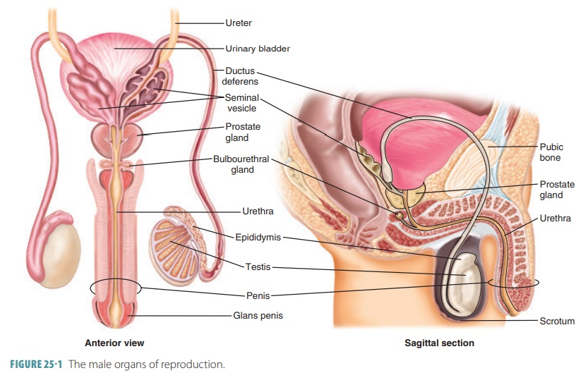
Urinary bladder receives a duct from the seminal vesicle to form ejaculatory duct that runs through the prostate and opens into urethra.
(e) Urethra receives the ducts of prostate gland and the bulbourethral gland (Cowper’s glands) a little ahead and runs through the penis to its external opening called urethral meatus.
(iii) The accessory glands of male reproductive system include
(a) A pair of seminal vesicles, a prostate gland and a pair of bulbourethral glands (Cowper’s glands).
(b) The secretion of all these glands is called seminal plasma.
(c) Seminal plasma contains fructose, calcium and some enzymes. It is to provide nutrition to the spermatozoa, while travelling through female reproductive tract.
(d) Seminal plasma along with sperms is called semen.
(e) Secretion of bulbourethral glands also helps in the lubrication of the penis.
(iv) External genitalia is the penis. It is made up of special erectile tissue that helps in erection of the penis. The enlarged tip of the penis is called glans penis. It is covered by a loose fold of skin called foreskin or prepuce.
3. Female reproductive system consists of a pair of ovaries, secondary sex organs, external genitalia and mammary glands.
(i) Ovaries are primary female sex organs which produce female gametes called ova and secrete the female sex hormones.
(a) These are located one on each side of the lower abdomen.
(b) It is almond-shaped, 2-4 cm in length, 1.5 cm in width.
(c) It is connected to the pelvic wall and uterus by ligaments.
(d) Each ovary is covered by a thin epithelium which encloses the ovarian stroma.
(e) Stroma is divided into two regions, i.e. peripheral cortex and inner medulla.
(ii) The female accessory ducts constitute oviducts (Fallopian tubes), uterus and vagina.
(iii) Each Fallopian tube is about 10-12 cm long and extends from the periphery of each ovary to the uterus.
Anatomy of the Male Reproductive System
Chapter: Anatomy and Physiology : Reproductive System
Anatomy and Structure of Scrotum, Testes, Penis, Male Duct System, Male Accessory Glands, Semen
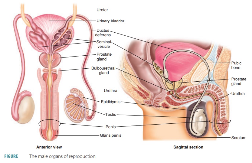
Anatomy of the Male Reproductive System
The structures of the male reproductive system include two epididymides, two ductus deferentia, two ejacula-tory ducts, the urethra, two seminal vesicles, the pros-tate gland, and two bulbourethral glands. Sperm cells are produced and maintained by the male reproduc-tive organs, which also transport these cells outside the body and secrete male sex hormones. The primary sex organs or gonads of the male consist of the two tes-tes, in which sperm cells and male sex hormones are formed. The accessory sex organs are the internal and external reproductive organs (FIGURE 25-1).
Scrotum
The male external reproductive organs consist of the scrotum and the penis. The scrotum consists of a flesh pouch of skin and subcutaneous tissue suspended below the perineum and anterior to the anus. The scro-tum encloses the testes. Sparse hairs cover the scrotum externally. Internally, the medial septum or raphe sub-divides it into two chambers, each enclosing a testis. The scrotum protects and controls the temperature of the testes, which is important for sex cell production.
When environmental temperatures are cold, the scrotum contracts and wrinkles, moving the testes closer to the pelvic cavity to absorb heat. When it is warmer outside, the scrotum relaxes and hangs loosely to ensure the testes are about 3°C lower than body temperature. This is better for the sperm cells to be produced and to survive. Viable sperm cannot be produced at normal core body temperature, which is 98.6°F (37°C).
The scrotum contains two sets of muscles that respond to temperature changes. Each dartos muscle is a smooth muscle layer in the superficial fascia that acts to wrinkle the scrotal skin. Bands of skeletal mus-cle arising from the internal oblique muscles of the body’s trunk are known as the cremaster muscles, which act to elevate the testes toward the body, to con-trol scrotal temperature.
Testes
The testes are oval-shaped structures, about 4 cm (1.5 inches) long and 2.5 cm (1 inch) wide, located within the cavity of the scrotum. Each testis is also enclosed in two tunics. The outer is the tunica vaginalis, which has two serous layers and is formed from a peritoneal outpocket. The inner tunic is the tunica albuginea and is a fibrous capsule. Thin septa extend inward from the tunica albuginea and divide the testis into approximately 250 lobules. Each wedge-shaped lobule contains one to four highly coiled seminiferous tubules that, when uncoiled, may reach 80 cm in length. It is here where sperm are actually formed. The seminiferous tubules are made up of a thickened stratified epithelium that surrounds a central lumen filled with fluid. Spermatogenic cells are found in larger columnar cells known as sustenocytes. These cells play a variety of roles in sperm formation. Sperm are generated continuously. The sustenocytes maintain the blood-testis barrier, support spermiogenesis, secrete inhibin hormone, and secrete androgen-binding protein.
A normal testis contains nearly one-half of a mile of seminiferous tubules. Each of these tubules forms a loop connected to a network of passageways known as the rete testis. Fifteen to 20 large efferent ductules connect the rete testis to the epididymis. This tube coils on the outer surface of the testis and becomes the ductus deferens (FIGURE 25-2). Three to five layers of myoid cells, which resemble smooth muscle cells, surround each seminiferous tubule. Rhythmic con-tractions of the myoid cells aid in squeezing sperm and testicular fluids through the seminiferous tubules and out of the testes. The rete testis receives sperm through a straight tubule formed by the seminiferous tubules of each lobule, which leads into the epididymis for the maturation of sperm. The epididymis wraps around the posterior external surface of each testis. Immature sperm pass through the head and body of the epidid-ymis to be stored in its “tail” portion until ejaculation.
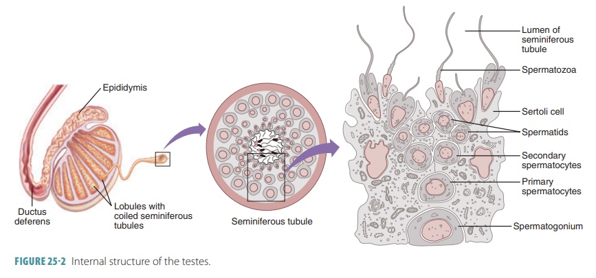
Interstitial endocrine cells, also known as Leydig cells, lie inside the soft connective tissue that surrounds the seminiferous tubules. They produce testosterone and less important types of androgens. These substances are secreted into the surround-ing interstitial fluid. The testes are supplied by long testicular arteries that branch from the abdominal aorta, superior to the pelvis. The testes are drained by the testicular veins, which arise from a network known as the pampiniform venous plexus. This network surrounds each testicular artery inside the scrotum, winding around it. In each pampiniform plexus, cooler venous blood absorbs heat from arterial blood. Therefore, this blood becomes cooler before entering the testes, which helps to keep the testes at their normal, cool, homeostatic temperature.
The testes are served by the sympathetic and para-sympathetic divisions of the autonomic nervous sys-tem. Forceful trauma to the testes transmits impulses, causing intense pain and nausea. In the testes, blood vessels, nerve fibers, lymphatic vessels, and the ductus deferens are enclosed by a connective tissue sheath. Together, these structures comprise the spermatic cord passing through the inguinal canal.
1. Explain the role of the scrotum in protection of the testes.
2. Describe the two major functions of the testes.
3. Identify the structures in which sperm are actually formed.
Penis
The penis is cylindrical in shape and conveys urine and semen through the urethra. When erect, it stiff-ens and enlarges, enabling insertion into the vagina during sexual intercourse. The penis is divided into three regions: the root, body, and glans or glans penis. The root of the penis is the fixed portion that attaches the penis to the body wall. At birth, skin that covers the penis is loose. It slides distally over the head, forming the foreskin or prepuce around the glans. The prepuce is often removed surgically soon after birth in a procedure called a circumcision. This practice is more common in the United States, where at least 65% of males are circumcised. In the rest of the world, about 30% of males experience this procedure. Many cultures are not familiar with circumcision, including some Hispanic cultures and many European and Asian cultures. Most males from Muslim and Jew-ish cultures are circumcised. It is widely believed that circumcision reduces risk of acquiring HIV or other reproductive system infections.

The dorsal and ventral surfaces of the penis are actually named in relation to the penis being erect, not flaccid. The shaft or body of the penis is the tubular, movable portion of the organ. It contains three columns of erectile tissue. It has two dorsal corpora cavernosa and one ventral corpus spongiosum (FIGURE 25 -3). The corpora cavernosa is the erectile tis-sue that is located on the anterior surface of the penis. The urethra is surrounded by the corpus spongiosum. Dense connective tissue surrounds each column in a capsule. The penis is enclosed by a layer of connective tissue, a thin layer of subcutaneous tissue, and skin. The erectile tissue of the penis contains many vascular spaces. These spaces fill with blood during sexual stimulation.
Male Duct System
When sperm are produced by the testes, they move out of the body via a system of ducts. These ducts are the epididymis, ductus deferens, ejaculatory duct, and urethra.
Epididymis
The cup-shaped epididymis can be felt through the skin of the scrotum. It is coiled and twisted to take up only a small amount of space, about 3.8 cm or 1.5 inches. The head of the epididymis contains effer-ent ductules and lies above the superior aspect of each testis. The body and tail of the epididymis are found on the posterolateral area of each testis.
The epididymis controls the composition of the fluid produced by the seminiferous tubules. It also absorbs and recycles damaged spermatozoa and absorbs cellular debris. The products of the break-down of enzymes are released into the surrounding interstitial fluids for pickup by the epididymal blood vessels. The epididymis also stores and protects sper-matozoa and facilitates their functional maturation.
Most epididymides are tightly coiled tubes about 6 m or 20 feet in length, connected to the posterior border of the testes. These tubes are also described as the ducts of the epididymides. They course upward to become the ductus deferens. In the duct mucosa, certain pseudostratified epithelial cells have stereocilia, which are long, nonmoving microvilli. Having a large surface area, the stereocilia can absorb extra testicular fluid and pass nutrients to the millions of sperm cells temporarily stored in the lumen.
Immature sperm cells are nonmotile when they reach the epididymis; therefore, rhythmic peristal-tic contractions move them through the duct as they mature. The surrounding fluid contains antimicrobial proteins such as defensins. The transfer of sperm from the testes and through the epididymides takes approx-imately 20 days. Once mature, sperm cells can move independently to fertilize egg cells, but usually do not actually “swim” until after ejaculation. This occurs from the epididymides and not the testes. The secretions of the seminal vesicles are discharged into the ejaculatory duct at emission. This is when peristaltic contractions are occurring in the ductus deferens, seminal vesicles, and prostate gland. These contractions are controlled by the sympathetic nervous system. Although sperm are normally stored in the epididymides for several months, longer storage results in them being phagocy-tized by the epithelial cells there.
Ductus Deferens and Ejaculatory Duct
The ductus deferentia, also called the vasa deferentia, are muscular tubes approximately 45 cm (18 inches) in length. Singularly, each ductus defer-ens is called a vas deferens. They each pass upward, as part of the spermatic cord, along the medial side of a testis, through the inguinal canal in the lower abdominal wall to enter the pelvic cavity. They end behind the urinary bladder, uniting just outside the prostate gland with the duct of a seminal vesicle. This forms an ejaculatory duct, which is a short structure passing through the prostate gland to empty into the urethra. The vas deferens is the duct that is altered when a male undergoes a vasectomy.
Each ductus deferens can easily be felt as it passes anterior to the pubic bone, looping medially over the ureter and descending along the posterior wall of the bladder. The terminus portion expands to form an ampulla, joining the duct of a seminal vesicle. The mucosa of the ductus deferens is, like the epidid-ymis, pseudostratified epithelium. It differs in that its muscular layer is very thick. During ejaculation, the smooth muscle of its walls creates peristaltic waves, quickly squeezing sperm forward into the urethra.
Male Accessory Glands
The male accessory glands consist of a pair of seminal glands and bulbourethral glands, plus a single prostate gland.
Seminal Glands
The seminal glands are also called seminal vesicles and are sac-like structures lying on the posterior bladder surface. They are approximately 5 cm long, attached to the ductus deferens near the base of the bladder. Each seminal vesicle is a tubular gland having a total uncoiled length of about 15 cm. However, these glands are normally coiled back on themselves, mak-ing their coiled size only 5–7 cm. During ejaculation, they are emptied by a thick layer of smooth muscle that contracts inside their fibrous capsules. They have glandular tissue linings that contribute nearly 60% of semen volume.
The seminal vesicles secrete a slightly alkaline fluid that is yellowish in color and viscous. This fluid helps to regulate the pH of the tubular contents as sperm cells travel to outside the body. The yellow color of seminal fluid comes from a pigment that becomes flu-orescent under ultraviolet light, a fact that is used for the investigation of certain crimes. Seminal fluid con-tains fructose, a monosaccharide that provides energy for sperm cells, as well as prostaglandins that stimu-late muscular contractions within the female repro-ductive organs. These contractions aid the movement of sperm cells toward the egg cell. The fluid from the seminal glands also contains citric acid and a coagu-lating enzyme known as vesiculase.
Remember that the duct of each seminal gland joins the duct of the ductus deferens on the same side, forming the ejaculatory duct. Here, seminal fluid mixes with sperm, entering the prostatic urethra simultaneously during ejaculation. Semen, therefore, is 70% made up by seminal gland secretions.
Prostate
The prostate gland surrounds the proximal portion of the urethra, slightly inferior to the urinary bladder. It is a chestnut- or doughnut-shaped, muscular struc-ture that is approximately 4-cm wide and 3-cm thick. It is surrounded by a thick connective tissue capsule and made up of 20–30 branched tubular glands with ducts that open into the urethra. These glands are embedded in a stroma, which is a mass of dense con-nective tissue and smooth muscle.
The prostatic smooth muscle contracts during ejaculation. Prostatic secretions are squeezed into the prostatic urethra through several ducts. The secretions consist of a milky fluid that is slightly acidic. Prostatic fluid enhances the motility of the sperm cells and helps neutralize the vagina’s highly acidic secretions. It makes up to one-third of the volume of the semen. Prostatic fluid contains citrate, which provides nutri-ents, prostate-specific antigen, and enzymes such as fibrinolysin, acid phosphatase, and hyaluronidase.
Bulbourethral Glands
The bulbourethral glands, also known as Cowper’s glands, are about 1 cm in diameter and lie inferior to the prostate gland surrounded by the external urethral sphincter muscle’s fibers. These glands have tubes with epithelial linings secreting a thick, clear mucous-like fluid as a response to sexual stimulation. The fluid lubricates the end of the penis to prepare for sexual intercourse, even though females secrete most of the lubricating fluid needed for sexual intercourse. The fluid from the bulbourethral glands also neutralizes any urine, which is acidic.
Semen
The milky white, slightly sticky fluid the male ure-thra conveys to outside of the body during ejacula-tion is known as semen. It is made up of sperm cells from the testes and secretions of the seminal vesicles, prostate gland, and bulbourethral glands. Semen has an alkaline pH of between 7.2 and 8.0, and includes prostaglandins and nutrients. It helps to neutralize the acidic environment of the male urethra and the female vagina. Under acidic conditions, the sperm “swim” more slowly than normal.
Between 2 and 5 mL of semen are released at one time, with between 20 and 150 million sperm/mL. How-ever, sperm only make up about 10% of the semen. Sperm cells begin to swim as they mix with accessory gland secretions. They acquire the ability to fertilize a female egg cell once they are inside the female reproductive tract in a process called capacitation, which is due to the weakening of the sperm cells’ acrosomal membranes.
Mature sperm do not contain significant amounts of stored nutrients or cytoplasm. Nearly all energy needed for sperm adenosine triphosphate synthe-sis is provided by the catabolism of fructose in sem-inal gland secretions. The prostaglandins in semen decrease viscosity in the femalecervix, stimulating reverse peristalsis. This speeds up the movement of sperm through the female reproductive tract. Semen also contains the hormone relaxin, various enzymes, ingredients that suppress the immune response in the female reproductive tract, antibiotics that destroy certain bacteria, and clotting factors. Just after ejacu-lation, the clotting factors coagulate the semen, which causes the sperm to stick to the vaginal walls of the female so they do not drain out of the vagina. Fibri-nolysin then liquefies the sticky mass, allowing the sperm to swim along their journey to the ovum.

(a) The part of oviduct closer to the ovary is funnel-shaped infundibulum.
(b) The edges of infundibulum possess finger-like projections called fimbriae, which help in collection of the ovum after ovulation.
(c) Infundibulum leads to a wider part of the oviduct called ampulla.
(d) Isthmus is the last part of the oviduct, which has a narrow lumen and it joins the uterus.
(iv) Uterus or womb is a pear-shaped muscular organ. It is attached to the pelvic wall and supported by ligaments.
(a) Wall of the uterus has three layers of tissue.
(b) Perimetrium is the outermost thin membranous layer, myometrium is the middle thick layer of smooth muscles and endometrium is the innermost glandular layer which lines the uterine cavity.
(c) Uterus opens into the vagina through a narrow cervix, its cavity is called cervical canal, which along with vagina forms birth canal.
(d) Endometrium layer undergoes cyclic changes during menstrual cycle.
(e) Smooth muscles in myometrium contract during parturition to deliver the baby.
(v) Vagina is a muscular tube-like structure that opens to the outside. It receives spermatozoa during insemination and serve as birth canal.
(vi) Female external genitalia include mons pubis, labia majora, labia minora, clitoris and hymen.
(a) Mons pubis is a cushion of fatty tissue covered by skin and pubic hair.
(b) Labia majora are fleshy folds of tissue which extend down from the mons pubis and surround the vaginal opening.
(c) Labia minora are paired folds of tissue under the labia majora.
(d) Hymen is a membrane that covers the opening of vagina partially. It gets ruptured during vigorous physical activities or during the first coitus.
(e) Clitoris is a tiny finger-like structure, which lies at the upper junction of the two labia minora above the urethral opening.
(vii) Mammary glands (breasts) are paired structures that contain glandular tissue and variable amount of fat.
(a) Glandular tissue of each mammary gland is divided into 15-20 mammary lobes containing the cluster of cells called alveoli.
(b) The cells of alveoli secrete milk, which is stored in the cavities (lumen) of alveoli.
(c) Alveoli open into mammary tubules. The tubules of each lobe join to form a mammary duct.
(d) Several mammary ducts join to form a wider mammary ampulla, which is connected to lactiferous duct through which milk is sucked out.
Anatomy of the Female Reproductive System
Chapter: Anatomy and Physiology: Reproductive System
Anatomy and Structure of the Female Reproductive System - Ovaries; Female Duct System: Uterine Tubes, Uterus, Uterine Supporting Structures, Layers of the Uterine Wall, Vagina ; External Genitalia ; Mammary Glands
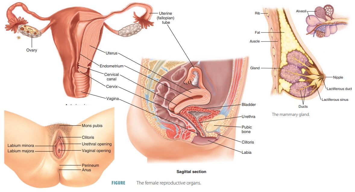
Anatomy of the Female Reproductive System
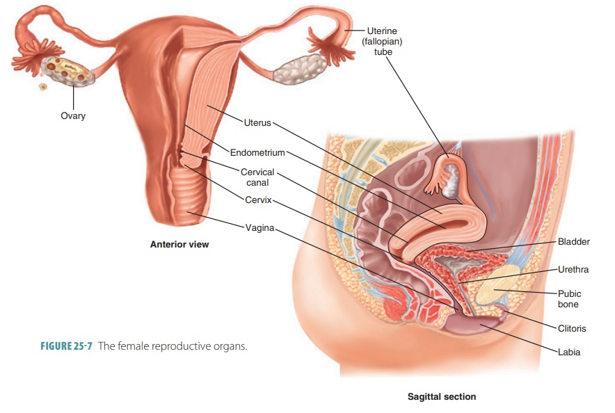
The anatomy of the female reproductive system is highly complex in comparison with that of the male and the female reproductive and urinary tracts are totally separated. The female reproductive organs pro-duce and maintain the egg cells or oocytes, which are the female sex cells. The organs also transport them to the site of fertilization, provide a strong environ-ment for the developing fetus, give birth to a fetus, and produce female sex hormones. The principal organs of the female reproductive system, besides the ovaries, are the uterine tubes, uterus, vagina, and the compo-nents of the external genitalia. The primary sex organs or gonads are the two ovaries, which reproduce female sex cells and sex hormones. The accessory sex organs are the internal and external reproductive organs (FIGURE 25-7). As in males, a variety of accessory glands releases secretions into the female reproductive tract. The female internal genitalia are primarily located in the pelvic cavity, and include the ovaries and duct sys-tem. The accessory ducts include the uterine tubes, uterus, and vagina.
Ovaries
The female gonads or ovaries are oval-shaped, solid structures about 3.5 cm long, 2 cm wide, and 1-cm thick. They lie in shallow depressions in the lateral pelvic cavity wall on either side of the uterus. The ova-ries are suspended by several ligaments in the perito-neal cavity, where the iliac blood vessels split into a “fork.” Each ovary is anchored medially to the uterus by an ovarian ligament and laterally to the pelvic wall by the suspensory ligament. Also, a mesovarium suspends each ovary in between these points. The mesovarium and suspensory ligament are part of a broad ligament, which folds over the uterus to sup-port the uterus, uterine tubes, and vagina. The ovarian ligaments are enclosed by the broad ligament.
The ovarian arteries serve the ovaries and are branches of the abdominal aorta. The ovaries are also served by the ovarian branch of the uterine arteries. To reach the ovaries, the ovarian blood vessels must travel through the mesovaria and suspensory ligaments.
Each ovary is externally surrounded by a fibrous tunica albuginea. This structure is then covered by a cuboidal epithelial cell layer that is known as the germinal epithelium. This epithelium is a continua-tion of the peritoneum. Each ovary additionally has an outer cortex enclosing the developing gametes. An inner medulla contains the primary blood vessels and nerves. However, the relative area of each region is not well defined.
Many small structures called ovarian follicles resemble sacs and are embedded in the cortex of each ovary, which is highly vascular and made of connective tissue. One oocyte is found in each folli-cle, encased in a variety of cells. If only a single layer is present, these are called follicle cells, but if more than one layer is present, they are called granulosa cells (FIGURE 25-8).
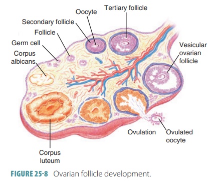
In women of childbearing age, one ripening fol-licle ejects its oocyte from an ovary every month in a process called ovulation. The ruptured follicle then changes its appearance, becoming a glandular struc-ture, the corpus luteum. This structure soon degener-ates. The surfaces of the ovaries show pits and scars in older women because they have released many oocytes over a lifetime.
Ovarian tissues consist of an inner medulla and an outer cortex. The medulla is made up of loose connective tissue with many blood and lymphatic vessels as well as nerve fibers. The cortex has more compact tissue with a granular appearance because of masses of ovarian follicles. The ovary’s free surface is covered with cuboidal epithelium above a layer of dense connective tissue. The almond-shaped ovaries perform three main functions: production of imma-ture female gametes called oocytes; secretion of female sex hormones, including estrogens and progestins; and secretion of inhibin, which is involved in the feed-back control of pituitary FSH production. The most common form of estrogen is estradiol, followed by estrone and estriol.
Before birth, a female fetus develops small cell groups in the outer ovarian cortex that form several million primordial follicles. Each follicle consists of a primary oocyte surrounded by follicular cells. The primary oocytes begin to undergo meiosis early in development, but then the process stops and does not restart until puberty. No new primordial fol-licles form after the initial ones form, and oocytes degenerate. Although several million oocytes form in the female embryo, only about 1 million remain at birth, with only 400,000 left at puberty. The ovary releases less than 400–500 oocytes during a female’s reproductive life.
Female Duct System
The female duct system has no or very little contact with the ovaries. The female reproductive system includes accessory structures, including two uterine tubes, a uterus, and a vagina.
Uterine Tubes
The uterine tubes, also called the fallopian tubes or oviducts, receive the ovulated oocytes from the ovaries and are each about 10 cm (4 inches) long. The uterine tubes are the sites where fertilization usually occurs. Each uterine tube empties into the superolateral area of the uterus via a constricted isthmus. As it curves around the ovary, each uter-ine tube’s distal end expands to form an ampulla. Near the ovaries, each tube expands into a funnel shaped infundibulum that partially encircles the ovary. Finger-like fimbriae surround its margin with one of the larger extensions connecting with the ovary.
The epithelium lining the uterine tube is com-posed of ciliated columnar epithelial cells, with scattered mucin-secreting cells. The mucosa is sur-rounded by concentric smooth muscle layers. The transport of oocytes involves a combination of cil-iary movement and peristaltic contractions in the uterine tube walls. Nonciliated mucosal cells have dense microvilli and produce secretions that keep oocytes as well as any present sperm nourished and moist. The uterine tubes are externally covered by peritoneum, supported by a short mesentery called the mesosalpinx. This structure is actually part of the broad ligament.
Uterus
If the secondary oocyte is fertilized to become a zygote, the uterus receives the developing embryo, sustaining its development. The uterus is hollow and muscular, shaped slightly like an inverted pear. Its size changes during pregnancy, from about 7.5 cm by 5 cm by 2.5 cm to much larger, able to hold the developing baby up until birth. At this point, it weighs 30–40 g. The uterus is located in the anterior pelvic cavity, superior to the vagina, usually bending over the urinary bladder. The uterine body is also called the corpus, the largest portion of the uterus. The fundus is the rounded portion of the corpus and is superior to the attachment of the uterine tubes. It ends at the constriction known as the isth-mus. The cervix is the inferior portion of the uterus, extending from the isthmus to the vagina. The cervix surrounds the cervical orifice, where the uterus opens to the vagina. The uterine wall is thick, with three layers.
The cervical canal communicates with the vagina through the external os and also with the uterine body cavity through the internal os. Cervical glands exist in the mucosa of the cervical canal and secrete mucus that fills the cervical canal and also covers the external os. This is believed to block bac-teria from spreading into the uterus from the vagina. In most times during the uterine cycle, this cervical mucus blocks sperm entry. However, it allows sperm to pass through at the midpoint of the cycle, which is when it becomes less viscous.
Uterine Supporting Structures
Additional supports of the uterus include the mesome-trium, laterally, and other ligaments. The cardinal lig-aments, also called lateral cervical ligaments, extend from the cervix and superior vagina to the lateral pelvic walls more inferiorly than the mesometrium. Two uterosacral ligaments secure the uterus to the sacrum, posteriorly. Fibrous round ligaments bind the uterus to the anterior body wall. They pass through the inguinal canals, anchoring in the labia majora’s subcutaneous tissue. Collectively, these ligaments allow the uterus to be quite movable, accommodating filling and emptying of the bladder and rectum.
Layers of the Uterine Wall
The three layers of the wall of the uterus are the perimetrium, myometrium, and endometrium. The perimetrium is the outer, incomplete, serous layer. The myometrium is the thick middle layer. It is made of interlaced smooth muscle bundles and contracts rhythmically during childbirth, expelling the baby from the uterus.
The endometrium is the mucosal lining, made of simple columnar epithelium above an underlying, thick lamina propria. Fertilization causes the embryo to implant into the endometrium for the entire preg-nancy. There are two chief layers or strata in the endometrium. The functional layer is the stratum functionalis, which changes based on ovarian hor-mone levels in the blood. This layer is shed during menstruation, about every 28 days. The basal layer is the stratum basalis, which is thinner. It forms a new stratum functionalis after menstruation. This layer does not respond to ovarian hormones. Many uterine glands in the endometrium change lengthwise, along with the endometrial thickness changes that occur.
The cyclic changes of the uterine endometrium are linked to its vascular supply. From the internal iliacs of the pelvis arise the uterine arteries. They ascend along the sides of the uterus, branching into the uterine wall. The branches split into several arcuate arteries inside the myometrium. These arteries continue as radial arteries into the endometrium. Here, straight arteries supply the stratum basalis, whereas spiral, coiled arter-ies supply the stratum functionalis. The spiral arteries degenerate and regenerate continuously. When they spasm, these actions cause the functionalis layer to be shed during the menstrual cycle. In the endometrium, the veins have thin walls. They form an extensive net-work with small amounts of sinusoidal enlargements.
Painful menstruation is known as dysmenorrhea. It may be caused by inflammation of the uterus, myo-metrial contractions commonly known as cramps, or conditions that involve nearby pelvic structures.
Vagina
The vagina is a thin-walled fibromuscular tube, about 8–10 cm (3–4 inches) in length, extending from the cervix to the outside of the body. It conveys uterine secretions, receives the erect penis during intercourse, and provides the open channel for offspring. The vagina extends up and back into the pelvic cavity and lies posterior to the urinary bladder and urethra but anterior to the rectum. The urethra is parallel to the course of the vagina anteriorly. The vagina is attached to these other structures by connective tissues.
The hymen is a thin membrane of connective tis-sue and epithelium that partially covers the vaginal orifice in females who have not had sexual inter-course. It has a central opening that allows uterine and vaginal secretions to pass to the outside of the body. The hymen is extremely vascular and may bleed when it stretches or ruptures during initial sexual inter-course. It can also be ruptured by insertion of tam-pons, sports activities, or pelvic examinations. In rare cases, it is tougher than normal and requires a surgical procedure for normal intercourse to occur.
The three major functions of the vagina are to serve as a passageway for the elimination of menstrual flu-ids, to receive the penis during sexual intercourse, and to hold the spermatozoa before their passage into the uterus. The vagina forms the interior portion of the birth canal, through which the fetus passes during delivery.
The vaginal wall has three layers:
■■ Inner mucosal layer (mucosa): Stratified squamous epithelium with no mucous glands. Dendritic cells act as antigen-presenting cells. They may be the route of HIV transmission from an infected male. This layer has no glands but is lubricated by the cervical mucous glands. It also has a mucosal transudate that leaks from the vaginal walls. Large amounts of glycogen are released by its epithelia, which are metabolized anaerobically by bacteria to form lactic acid. Therefore, the pH is very acidic, which helps to fight infections but is harmful to sperm. Because this fluid is alkaline instead of acidic in adolescent girls, they are predisposed to STIs if they are sexually active.
■■ Middle muscular layer (muscularis): Mostly smooth muscle fibers; helps to close the vaginal opening.
■■ Outer fibrous layer (adventitia): Dense connective tissue and elastic fibers.
The vaginal fornix is a recess produced at the upper end of the vaginal canal, which loosely surrounds the cervix. The posterior fornix is much deeper than the lateral and anterior fornices. The lumen of the vagina is basically small, and its posterior and anterior walls touch each other, except where the cervix keeps it open. During sexual intercourse and childbirth, the vagina can stretch considerably. However, ischial spines and the sacrospinous ligaments limit its lateral distention. When various microorganisms cause inflammation and infection of the vagina, it is known as vaginitis.
1. Describe the internal genitalia of females.
2. Explain the roles of the ovaries.
3. Describe the three layers of the uterine wall.
4. Name the suspensory ligaments that support the uterus and hold it in place.
5. Describe normal and abnormal locations where fertilization may occur.
External Genitalia
The external accessory organs of the female reproduc-tive system include the mons pubis, labia majora, labia minora, clitoris, and vestibular glands (FIGURE 25-9).They surround the openings of the urethra and vagina, composing the vulva or pudendum. The mons pubis is a rounded area made of fatty tissue that overlies the pubic symphysis. This area becomes covered with pubic hair after puberty.
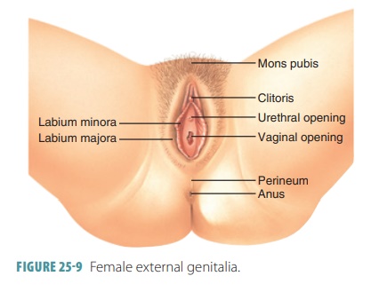
The labia majora enclose and protect the other external reproductive organs. They are made up of rounded folds of adipose tissue and thin smooth mus-cle covered by skin and hair. They lie close together, with a cleft that includes the urethral and vaginal openings separating the labia longitudinally. The labia majora are analogous to the male scrotum and enclose the labia minora.
The labia minora are flattened, hairless longitudi-nal folds composed of connective tissue. They contain the external openings of the urethra and vagina. They have a rich blood supply, and therefore a pinkish appear-ance. They merge posteriorly with the labia majora to form a ridge called the fourchette. Anteriorly, they converge to form the hood-like covering of the clitoris.
The clitoris projects from the anterior end of the vulva between the labia minora. It is usually about 2 cm in length and 0.5 cm in diameter. It corresponds to the penis in males, with a similar structure. It is made up of two columns of erectile tissue called the corpora cavernosa and forms a glans at its anterior end that has many sensory nerve fibers. The exposed portion is called the glans of the clitoris and the hooded fold is called the prepuce of the clitoris. The clitoris has a rich innervation of sensory nerve endings and swells with blood, becom-ing erect during tactile stimulation and sexual arousal.
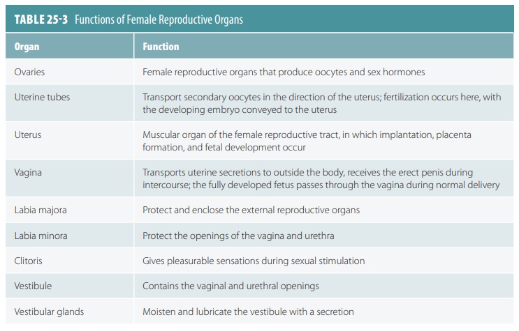
The labia minora encloses the vestibule, into which the vagina opens posteriorly. The urethra opens into the vestibule in the midline, about 2.5 cm posterior to the glans of the clitoris. One pea-sized vestibular gland lies on each side of the vaginal opening, which is similar to the bulbourethral glands of males. They release mucus into the vestibule, moist-ening and lubricating it for intercourse. Under the vestibule’s mucosa, on either side, is a mass of vascular erectile tissue called the vestibular bulb. These bulbs are similar to the single penile bulb and corpus spon-giosum in males. They engorge with blood during sex-ual stimulation, helping the vagina to grip the penis and causing the urethral orifice to shut. This prevents semen and bacteria from moving superiorly into the bladder as intercourse occurs. TABLE 25-3 summarizes the functions of the female reproductive organs.
Mammary Glands
The mammary glands are specialized to secrete milk after pregnancy. They are located in the subcutane-ous tissue of the anterior thorax within the breasts. The breasts are above the pectoralis major muscles, extending from the second to the sixth ribs, from the sternum to the axillae. They lie within the superficial fascia, also known as the hypodermis. The subcutane-ous tissue of a pectoral fat pad, which is deep to the skin of the chest, contains each mammary gland.
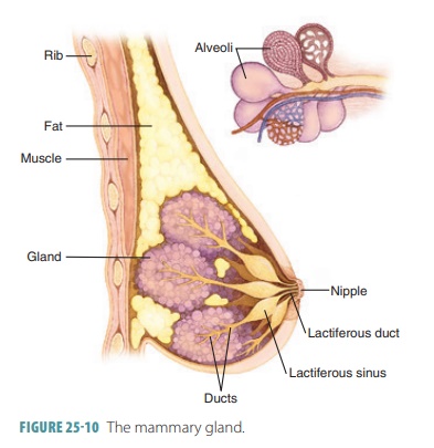
Just below the center of each breast is an areola, which is a ring of pigmented skin. The areola is slightly bumpy because of large sebaceous glands and produces sebum to reduce cracking and chapping of the nipple, which is located near the tip of each breast within the areola surrounding it (FIGURE 25-10). Smooth muscle fibers in the areola and nipple are controlled by the autonomic nervous system. This can cause the nipple to become erect when it is stimulated by touch or cold temperatures. Although present in males, the mam-mary glands have no function.
Each mammary gland is a modified sweat gland made up of 15–25 lobes that contain alveolar glands and an alveolar duct, which leads to a lactiferous duct. This leads to the nipple. The lobes are separated by dense adipose and connective tissues. Dense suspensory ligaments extend inward to help support the weight of the breast. Lobules are smaller units inside the lobes. They contain glandular alveoli, which produce milk during lactation. Milk is passed from these compound alveolar glands into lactiferous ducts, which open to the outside of the nipple. Each duct, just below the are-ola, has a dilated lactiferous sinus, where milk accumu-lates during nursing. As female children reach puberty, their mammary glands develop because of ovarian hormones. The alveolar glands and ducts enlarge. Fatty tissue deposits around and within the breasts.
1. Describe the structures of the external genitalia.
2. Explain the components of the mammary glands.
3. Identify risk factors for breast cancer.
Ovary – Female Reproductive System
The biological system made up of all the anatomical organs involved in sexual reproduction is known as an organism’s reproductive system, also referred to as the genital system. Numerous inanimate materials, including fluids, hormones, and pheromones, are crucial reproductive system add-ons. The sexes of distinct species frequently differ significantly from one another, unlike other organ systems. Due to these variations, two people’s genetic makeup can be combined, potentially resulting in kids with higher genetic fitness.
Human Ovary
The female reproductive organ known as the ovary houses an egg known as an ovum. After being discharged, this continues its journey through the fallopian tube and enters the uterus, where sperm may fertilize it. An ovary is located on either side of the body. The ovaries also release hormones that are important for fertilization and the menstrual cycle. Beginning in the prenatal phase and continuing into menopause, the ovary goes through a number of stages. It is frequently categorized as an endocrine gland because of the hormones it metabolizes.
Ovary Structure
The term “ovaries” refers to the female gonads. The ovarian fossa, which runs parallel to the uterus’ lateral wall, is where each ovary can be found. The area in front of the ureter and internal iliac artery, which is encircled by the external iliac artery, appears to be the ovarian fossa. This area is roughly 4 cm by 3 cm by 2 cm in size. The outer cortex and inner medulla of the ovaries, which are enclosed by a capsule, are seen in the anatomy of the ovary. A capsule called the tunica albuginea is constructed of thick connective tissue.
Each menstrual cycle, one of the two ovaries experiences ovulation, which results in the release of an egg.
The portion of the ovary closest to the fallopian tube is connected to the tube by the infundibulopelvic ligament, while the portion on the other side is joined to the uterus by the ovarian ligament. One of the additional tissues and structures of the ovaries is the hilum.

Structure of Ovary in Human
- Ligaments: The ovary’s slice reveals that the ovaries are joined by a fibrous ligament known as the ovarian ligament and are situated on either side of the uterus inside the peritoneal cavity. The ovary suspensory ligament, which seems to be a posterior extension of the uterus’ broad ligament, attaches the exposed ovaries to the body wall despite the fact that they are located in the peritoneal cavity. The part of the uterus’ thick ligament that covers the ovary is called the mesovarium. The ovarian pedicle is made up of fragments of the ovarian ligament, mesovarium, and blood vessels.
- Microanatomy: Simple cuboidal-to-columnar-shaped mesothelium makes up the membrane that covers the top of the ovaries, known as the germinal epithelium. The ovary’s internal architecture. Claims that the ovarian cortex is the outer layer, with the stroma in between the ovarian follicles. The follicles contain the cumulus oophorus, membrana granulosa (and its granulosa cells), zona pellucida, corona radiata, and primary oocyte. Theca of follicle, antrum, and liquid folliculin is frequently found in the follicle. The inner lining is called the ovarian medulla. Although it might be challenging to tell the cortex from the medulla, follicles are almost never found there. The ovary’s surface epithelium gives birth to follicular cells, which are flat epithelial cells. They are surrounded by granulosa cells, which have changed from flat to cuboidal and multiply to form a stratified epithelium. The ovary has lymphatic and blood arteries as well.
Functions
A child’s ovary starts to secrete more hormones when they approach puberty. The hormones cause secondary sex characteristics to manifest. The structure and function of the ovary alter beginning during puberty. The ovaries are important for fertility and pregnancy because they have the capacity to regulate hormones. Once egg cells (oocytes) are removed from the Fallopian tube, several feedback mechanisms activate the endocrine system, causing hormone levels to change. These feedback systems are under the direction of the hypothalamus and pituitary gland. The pituitary gland receives information from the hypothalamus.
In addition, the pituitary gland distributes hormones into the ovaries. In response to this signaling, the ovaries release their own hormones.
- Gamete Production: The ovaries are the site of the regular production and release of female gametes, also known as egg cells. The ovarian follicles, which are filled with fluid, are where the immature egg cells, also known as oocytes, mature. In the majority of cases, only one oocyte matures at a time, however, it is possible for others to mature concurrently. According to their stage of development, follicles include different kinds and numbers of cells, and the size of a follicle corresponds to the stage of oocyte growth. The pituitary gland releases luteinizing hormone when the oocyte has finished maturing in the ovary, which triggers the release of the egg by follicular rupture, or ovulation.
- Hormone Secretion: The mature ovaries secrete the hormones estrogen, inhibin, androgen, and progestogen. Up until menopause, 50% of a woman’s testosterone is found in the ovaries; beyond that, it is directly discharged into the circulation. DHEA and androstenedione, which are adrenal pre-androgens, are converted by other parts of the body into testosterone to make up the remaining 50% of testosterone in the blood. During puberty, estrogen plays a role in the development of secondary sex traits in females as well as the maturity and maintenance of fully developed reproductive organs.
- Ovarian Aging: As a woman matures, her ability to reproduce lessens, resulting in menopause. This drop is associated with a decrease in the number of ovarian follicles. At birth, the human ovary contains roughly 1 million oocytes, but only 500 (or about 0.05 percent) of these ovulate, with the rest being destroyed. As women get older, their ovarian reserves tend to decline at a constant rate, approaching total depletion around age 52. Age-related declines in ovarian reserve and fertility are accompanied by an increase in pregnancy failure and meiotic mistakes, which lead to chromosomally abnormal pregnancies. Ovarian reserve and fertility peak between the ages of 20 and 30. Around the age of 45, the menstrual cycle begins to change, and the follicular pool tends to significantly decrease. We don’t know what causes ovarian aging. Aging variability is influenced by environmental circumstances, dietary habits, and genetic factors.
Ovulation
A woman’s menstrual cycle typically lasts around 28 days, though this varies widely from woman to woman. When the endometrial lining sheds, the first day of the menstrual cycle is regarded as the first day of menstruation.
Every woman’s monthly cycle has specific times when significant things take place, making her fertile and may be able to produce a kid. The cycle begins with the follicular phase, which is followed by ovulation. The luteal phase continues for the remainder of the period till the cycle restarts.
Each of these procedures is triggered by hormones produced by the anterior lobe of the pituitary gland under the direction of the hypothalamus. Two particularly significant hormones for the menstrual cycle are luteinizing hormone (LH) and follicle-stimulating hormone (FSH).
Follicular phase
- The follicular phase sometimes referred to as the proliferative phase, lasts from the start of menstruation through ovulation and is the time when the follicles in the ovaries mature.
- Both cumulus oophorous granulosa cells and corona radiate must assist the egg for ovulation to occur successfully. These cells multiply and become mucous during cumulus expansion, secreting a fluid rich in hyaluronic acid. It has been demonstrated that this material must combine with the cell network to create a sticky matrix surrounding the egg in order for fertilization to occur.
- Additionally, there are more cumulus cells, which results in more antrum fluid being produced. As a result, the follicle enlarges to a diameter of over 20 millimetres (mm), generating the blister, a protrusion at the surface of the ovary.
- Estrogen levels peak near the conclusion of this period, which triggers the hormonal shifts required to start ovulation.
Ovulation
- The pituitary gland releases an abrupt increase in LH and FSH hormones as a result of the estrogen peak.
- Before the follicle bursts and releases the egg from the ovary through the oviduct, this normally lasts for 1 to 2 days. Due to LH, which makes the follicle secrete proteolytic enzymes that weaken the tissue surrounding the follicle’s blister and finally form a hole known as the stigma, the egg is released.
- The cumulus-oocyte complex, which is the name given to the egg encircled by cumulus cells, then goes into the peritoneal cavity and attaches to the fimbriae at the end of the fallopian tube. The egg gently moves towards the uterus while being propelled along the tube by cilia.
- The egg goes through meiosis I at the same time to split into two distinct cells: a polar body that is dormant and one that contains the cytoplasm.
- The egg then enters Meiosis II, although it does not finish since it continues in the metaphase until fertilization. The egg will deteriorate if it is not fertilized within 24 hours. The endometrial glands are still non-secretory, and the functional, uterine mucous membrane, has reached its maximal size at this time.
Luteal phase
- The follicle reaches the end of its life during this phase. The follicle collapses in on itself without the egg to create the corpus luteum, which can secrete the hormones progesterone and estrogen.
- These hormones cause the endometrial glands to produce proliferative endometrium, which is where the embryo will develop if implantation does place.
- Because progesterone is present, the basal body temperature slightly rises. The paracrine function of the corpus luteum keeps the endometrium healthy for the rest of the menstrual cycle.
- When the endometrium breaks down into scar tissue and menstruation starts, the cycle is over and the follicular phase officially begins.
FAQs on Female Reproductive System
Question 1: How Can Your Ovaries Be Affected by Certain Factors?
Answer:
The following list includes the elements that can affect your ovaries:-
- Ovulation abnormalities are issues with the ovaries’ ability to release eggs.
- Uterine or cervical malformations, as well as fibroids or polyps in the uterus.
- Damage to or obstruction of the fallopian tube, which is frequently brought on by inflammatory pelvic illness.
Question 2: What is the cervix?
Answer:
The cervix is the lower, more restricted, and bifurcated section of the uterus. The lower vaginal portion projects into the anterior wall of the vagina, while the upper supravaginal portion interacts with the uterus’s body. The birth canal is made by the vagina and the cervical canal.
Question 3: What are the uterus’ three layers?
Answer:
The uterus’s outer, serous layer, which develops from the peritoneum, is covered by this layer. The middle muscular layer, or myometrium, follows. It is formed of smooth muscle fibres and is the thickest layer. The endometrium or inner mucus layer, a glandular layer, is the third layer.
Question 4: What are the ovary’s purposes?
Answer:
Endocrine and gametogenic functions are both carried out by female ovaries. The release of female sex hormones including oestrogen and progesterone is an endocrine function. The development and release of the egg or ovum for reproduction is the gametogenic function.
Question 5: What is an Ovary?
Answer:
One of two female glands that produce the feminine hormones progesterone and oestrogen as well as the eggs. These hormones are crucial for the development of the breasts, body form, and body hair in women.
Question 6: What is secreted by the corpus luteum?
Answer:
Progesterone is the main hormone secreted by the corpus luteum, however it also makes inhibin A and estradiol. The corpus luteum will eventually regress in the absence of fertilisation.
1. Male Reproductive System – Structure, Organs, Functions
All living creatures increase or duplicate and produce posterity of a comparable kind. Propagation is a fundamental interaction between the presence of animal groups and the continuation of life. Propagation is the cycle by which all life forms duplicate in number and increment their populace.
Male Reproductive System

- The male conceptive framework comprises organs that produce and transport the male microbe cell or gamete, male chemical testosterone, and the organs which work with the release of male microorganism cells into the female regenerative framework for preparation.
- The male gamete is the sperm which is a minuscule body containing the hereditary material and they have a long tail for motility to assist them with arriving at the female microorganism cell for treatment.
- The vitally regenerative organ in guys is a couple of testicles.
- They produce the male sex cells called sperms and furthermore produce the male sex chemical testosterone.
- The framework comprises a few outside organs like the penis, scrotum, and testicles and inside organs like the urethra, prostate, and original vesicles.
- The Penis has a root that is associated with the designs of both the pelvic bones and lower stomach (the shaft’s noticeable part) organs that has a cone molded end.
- The urethra opening is the channel that conveys semen and pee and lies at the tip of the penis. The foundation of the penis is known as Corona.
- The vast majority of the male conceptive framework is situated beyond your stomach hole or pelvis. The outer pieces of the male conceptive framework incorporate the penis, the scrotum, and the gonads.
Penis
The penis is the male organ for sex. It has three sections they are ;
- The root – This is the piece of the penis that appends to the mass of your midsection.
- The body or shaft – Shaped like a cylinder or chamber, the body of the penis is comprised of three inward chambers. Inside these chambers, there’s an exceptional, wipe-like erectile tissue that contains a great many enormous spaces that load up with blood when you’re physically stirred. As the penis loads up with blood, it becomes inflexible and erect, which considers entrance during sex. The skin of the penis is free and flexible, considering changes in penis size during an erection.
- The glans – This is the cone-formed finish of the penis. The glans, which are likewise called the top of the penis, are covered with a free layer of skin called a prepuce. This skin is here and there eliminated in a methodology called circumcision.
The kickoff of the urethra — the cylinder that transports both semen and pee out of the body — is situated at the tip of the glans penis. The penis likewise contains numerous delicate sensitive spots.
Testicles
A couple of testicles are available in people. Testicles are available external the body in a pocket called the scrotum. They are oval bodies, around 4 to 5 cm long and 2 to 3 cm wide. For the most part, the left testis hangs somewhat lower than the right one.
The two essential elements of tests are as per the following:
- Creating sperms or spermatogenesis – a transporter of man’s qualities.
- Creating testosterone – a male sex chemical.
- Every testis contains around 250 testicular lobules or compartments.
Sperms are created in the seminiferous tubules. Seminiferous tubules are lined by two sorts of cells:
- Sertoli cells – They give sustenance to microbe cells.
- Spermatogonia or male microbe cells – They go through spermatogenesis to deliver sperm.
- Ley dig cells or interstitial cells are available external to the seminiferous tubules in the interstitial spaces. They emit male sex chemicals or androgens, for example, testosterone.
Scrotum
- This is the free pocket-like sac of skin that hangs behind and underneath the penis. It contains the balls (likewise called testicles), as well as many nerves and veins.
- The scrotum goes about as an “environment control framework” for the testicles. For ordinary sperm improvement, the testicles should be at a temperature marginally cooler than the internal heat level.
- Exceptional muscles in the mass of the scrotum permit it to contract and unwind, drawing the balls nearer to the body for warmth or farther away from the body to cool the temperature.
Male Sex Accessory Organs
- Rete testis, vasa efferentia , epididymis, and vas deferens are male sex frill pipes. Seminiferous tubules open into rete testis, which prompts vasa efferentia. Vasa efferentia opens into the epididymis and epididymis prompts vas deferens.
- It prompts the ejaculatory conduit alongside a channel from the fundamental vesicle. Sperms mature in these conduits. Sperms are put away and shipped through these pipes.
Epididymis
The epididymis is a long, looped tube that lays on the rear of every gonad. It transports and stores sperm cells that are created in the testicles. It likewise is the occupation of the epididymis to carry the sperm to development, since the sperm that rise up out of the testicles are youthful and unequipped for treatment. During sexual excitement, withdrawals force the sperm into the vas deferens.
Urethra
It is a cylinder-like construction that interfaces the urinary bladder to the urinary meatus. In guys, the urethra goes through the penis and is for the most part engaged with two principal capabilities: The Prostate exists underneath the bladder and covers the urethra. The prostate becomes bigger with age. In the event that the prostate develops excessively, it can impede the pee course through the urethra and be answerable for a few urinary side effects. This area is remembered for the urinary lot that takes pee from the bladder where semen is discharged.
Vas deferens
The vas deferens is a long, solid cylinder that moves from the epididymis into the pelvic hole, to simply behind the bladder. The vas deferens transports mature sperm to the urethra, the cylinder that conveys pee or sperm beyond the body, in anticipation of discharge.
Ejaculatory conduits
These are framed by the combination of the vas deferens and the fundamental vesicles (see underneath). The ejaculatory conduits void into the urethra.
Male Sex Embellishment Organs
The male embellishment organs incorporate original vesicles, prostate, and bulbourethral organs. These organs emit liquids that enter the urethra.
Fundamental Vesicles
The matched original vesicles are saccular organs back to the urinary bladder. Every organ has a short channel that gets together with the ductus deferens at the ampulla to frame an ejaculatory conduit, which then discharges into the urethra. The liquid from the fundamental vesicles is gooey and contains fructose, which gives an energy source to the sperm; prostaglandins, which add to the versatility and feasibility of the sperm; and proteins that cause slight coagulation responses in the semen after discharge.
Prostate
The prostate organ is a firm, thick design that is found only second rate compared to the urinary bladder. Various short channels from the substance of the prostate organ void into the prostatic urethra. The emissions of the prostate are flimsy, smooth-hued, and soluble. The capability to upgrade the motility of the sperm.
Bulbourethral Glands or Cowper organs
The matched bulbourethral organs are little, about the size of a pea, and situated close to the foundation of the penis. A short channel from every organ enters the proximal finish of the penile urethra. In light of sexual excitement, the bulbourethral organs discharge a basic bodily fluid-like liquid. This liquid kills the corrosiveness of the pee buildup in the urethra, assists with killing the causticity of the vagina, and gives a grease to the tip of the penis during intercourse.
Conceptual Question
Question 1: Make sense of the capability of the male regenerative framework?
Answer:
The entire male conceptive structure is dependent upon synthetic substances, which are engineered materials that control the development of many kinds of cells or organs. The fundamental synthetic substances related to the male regenerative structure are follicle fortifying compound, luteinizing compound, and testosterone.
Question 2: Explain about main reproductive organs in the male reproductive system?
Answer:
The super-regenerative organ in guys is a couple of testicles. They are available in scrotal sacs outside the body and contain seminiferous tubules as the primary and practical unit.,Male sex cells, sperms, are delivered by seminiferous tubules and mature in the epididymis. Leydig cells or interstitial cells in the middle of between the seminiferous tubules emit chemical testosterone.
Question 3: Explain about the penis?
Answer:
The penis is the male copulatory organ and a tube-shaped pendant organ found foremost to the scrotum and capabilities to move sperm to the vagina. The penis comprises three segments of erectile tissue that are enveloped by connective tissue and covered with skin.
Question 4: What are male sex accessory glands?
Answer:
The male frill organs incorporate fundamental vesicles, prostate, and bulbourethral organs. A couple of original vesicles are available. The Seminal vesicles are available over the prostate, connected with the vas deferens to make the ejaculatory conduits that move through the prostate. The male frill organs create a liquid that sustains the sperm. It is called fundamental plasma.
Question 5: What are the functions of testes?
Answer:
Testicles are a couple of egg-formed organs that demonstrate the scrotum, outwardly of the body. Their capability is To deliver male gametes for example the sperms and To deliver a male regenerative chemical called testosterone which is liable for creating sperms as well as optional sexual qualities in guys.
2. Female Reproductive System – Diagram, Functions, Organs
The female reproductive system is made up of the internal and external sex organs which play an important part in reproduction. In humans, the female reproductive system matures to maturity at puberty in order to create gametes and carry a fetus to term. The female reproductive system is immature at birth. The vagina, uterus, fallopian tubes, and ovaries are the internal sex organs.
Female Reproductive System
The female reproductive system includes both the primary and secondary sex organs as essential parts. The primary sex organs in females are two ovaries, which also emit female sex hormones including progesterone and estrogen in addition to producing ova or eggs. The uterus, fallopian tubes, cervix, and vagina are the secondary sex organs. The labia minora, labia majora, and clitoris make up the external genitalia. The mammary glands are also significant glands and play an important role in the female reproductive system. These components together help in the ovulation, fertilization, labor, and delivery processes, as well as child care.
Labeled Diagram of Female Reproductive System

Anatomy of the Female Reproductive System
The female reproductive system was designed to serve many functions. It produces ova, or egg cells, which are necessary for reproduction. The apparatus is set up to convey the ova to the area where fertilization will take place. In the fallopian tubes, the egg and sperm are fertilized. The next step for fertilized eggs is to implant in the uterine walls and start the phases of pregnancy. The female reproductive system also contributes to the generation of female sex hormones, which help to keep the reproductive cycle regular, in addition to all of the previously mentioned functions.
Ovaries
The primary female sex organs that create the female gamete and other hormones are the ovaries. One of these organs is located on either side of the lower abdomen. Each ovary is between 2 and 4 cm long, and ligaments connect them to the uterus and pelvic wall. The ovary is separated into two zones—the outer cortex and the inner medulla—and is covered by a thin layer of epithelium that encloses the ovarian stroma.
Numerous ovarian follicles in various developmental stages make up the cortex. The primary component of the female reproductive system is referred to as the ovarian follicle. Ampulla, isthmus, and infundibulum are the three anatomical regions that make up each oviduct.
Uterus
It is a muscular female reproductive organ with an inverted pear form. There are three layers that make up the uterus’s walls: an inside glandular layer, a middle thick layer, and an outside thin layer. Ligaments linked to the pelvic wall, which opens into the vagina from a thin cervix, support these three layers. The birth canal is made by the vagina and the cervical canal. A muscular tube called the vagina extends from the uterus’ lower end to the outside.
Fallopian Tubes
A pair of muscular, funnel-shaped structures called fallopian tubes run from the right and left superior corners of the uterus to the margin of the ovaries. The eggs discharged from the ovaries are picked up by these tubes, which are encased in tiny projections called fimbriae, which then transport them to the infundibulum where they supply the uterus. Cilia on each fallopian tube serve the purpose of transporting the ovum to the uterus.
Vagina
The muscular and elastic tube that links the cervix to the outside body is called the vagina. It serves as the penis’s container during sexual activity and transports sperm to the uterus and fallopian tubes. By enlarging to allow the delivery of the fetus during childbirth, it also serves as a birth canal.
Cervix
The uterine cervix, a tubular structure that connects the vagina and uterine cavity, serves as a passage between the two. At the cervical os, the inferior cervix opens into the upper vagina. The cervix’s lining that extends into the vagina is referred to as the ectocervix and is made up of stratified squamous epithelium. The endocervix, which is made of columnar epithelium, lines the interior of the cervical canal. The transformation zone is the area where the ecto- and endocervix converge and is distinguished by the change from columnar to squamous epithelium.
Labia Minora
The smaller lips are referred to as labia minora. They are a pair of very small cutaneous folds that descend from the clitoris. The anterior folds of the labia minora that encircle the clitoris create the clitoral hood and frenulum of the clitoris. The limits of the vulva vestibule are then formed by the labia minora deviating downward and obliquely.
Labia Majora
The lateral and longitudinal margins of the vulval clefts are defined by the labia majora, a prominent pair of cutaneous skin folds. The clitoris, labia minora, vulva vestibule, Bartholin’s glands, vestibular bulbs, Skene’s glands, vaginal entrance, and urethra are all protected by the folds they produce.
Clitoris
The clitoris is an erectile organ that resembles the male penis. It is made up of two corpora cavernosa, the glans, and prepuce. The female homologue of the penis, the clitoris is situated below the mons pubis. The clitoris is made up of two erectile vascular tissue corpora cavernosa that are adjacent to each other and encircled by a fibro collagenous sheath. A central septum that is incomplete partially separates the two corpora.
Ovulation
The process of releasing the eggs from the ovaries is called ovulation. When the follicle is fully developed and reaches its maximum size, this process occurs together with the buildup of fluids inside the follicle without a noticeably increased rise in pressure. The stigma, also known as the macula pellucida, first emerges jutting outward as a transparent cone area when the follicle grows out. Later, the stigma experiences localized changes in color, integrity, and translucency. Before ovulation, estrogen hormone release peaks at its highest level. At the site of the stigma, ovulation takes place after the luteinizing hormone surge. This increase is necessary for ovulation.
Menstrual Cycle
After reaching puberty, every month during a process known as the menstrual cycle, all females generate mature egg cells. A developed egg is released from an ovary during this time and goes to the uterus. If the egg in the uterus is not fertilized, the uterine lining sheds and a new cycle starts. A menstrual cycle typically lasts 28 days, however, it’s possible for them to last up to 35 days in some people or as little as 21 days in others. The endocrine system regulates the entire menstrual cycle, and the hormones FSH, LH, estrogen, and progesterone are involved. The pituitary gland produces both the FSH and LH hormones, whereas the ovaries generate estrogen and progesterone.

Except during pregnancy, menstrual cycles happen every month from the time of puberty until the age of 45 to 55. The release of mature eggs and hormone synthesis by the ovaries both decrease after the age of 55. Gradually, the menstrual cycle ends, making it impossible for the woman to get pregnant.
FAQs on Female Reproductive System
Q: What is the female reproductive system?
Answer:
The body system which is responsible for creating female gametes, often known as eggs or ova, as well as some sex hormones and keeping fertilized eggs alive until they mature into fetuses and are prepared for delivery is known as Female Reproductive System. The primary and accessory sex organs are integral components of the female reproductive system.
Q: Write the functions of the ovary.
Answer:
Endocrine and gametogenic functions are both carried out by female ovaries. The release of female sex hormones including estrogen and progesterone is an endocrine function. The development and release of the egg or ovum for reproduction is the gametogenic function.
Q: What is the cervix?
Answer:
The cervix is the lower, more restricted, and bifurcated section of the uterus. The lower vaginal portion projects into the anterior wall of the vagina, while the upper supravaginal portion interacts with the uterus’s body. The birth canal is made by the vagina and the cervical canal.
Q: What are fallopian tubes?
Answer:
A pair of muscular, funnel-shaped structures in the female reproductive system are called fallopian tubes. They run from the right and left superior corners of the uterus to the margin of the ovaries.
Physiology of the Female Reproductive System
Chapter: Anatomy and Physiology : Reproductive System
Physiology and Function of the Female Reproductive System : Oogenesis, Ovarian Cycle, Follicular Phase, Ovulation, Luteal Phase, Hormonal Regulation of the Ovarian Cycle, Uterine Cycle, Estrogen, Progesterone, and Female Reproductive Function, Female Sexual Response
Physiology of the Female Reproductive System
Females release egg cells only from puberty to meno-pause, which occurs on average at around age 51. Although today we know that egg stem cells continue to survive throughout life, it has not yet been proven that the cells are viable for reproduction. Physiology of the female reproductive system includes the pro-cesses of oogenesis, the ovarian cycle, hormonal reg-ulation, the uterine or menstrual cycle, and the female sexual response.
Oogenesis
Oogenesis is the process of egg cell formation, pro-ducing female sex cells. During the fetal period, the diploid stem cells of the ovaries or oogonia multiply quickly via mitosis. Eventually, primordial follicles appear, whereas the oogonia change into primary oocytes. They are surrounded by one layer of flat-tened follicle cells. The first meiotic division is begun by the primary oocytes. However, they stall in the late part of prophase I and do not complete their division.
At birth, a female infant is believed to have a cer-tain finite amount of primary oocytes. Although there were seven million oocytes originally, at birth about one million have survived programmed death. They are located in the cortical region of each immature ovary. By puberty, approximately 300,000 oocytes remain. Primordial follicles change into an enlarging collection of primary follicles over time. This process starts during the fetal period and continues until there are no more primordial follicles. At this time, meno-pause begins. In rare conditions, menopause occurs before age 40 and is known as premature menopause.
Oogenesis in the ovaries at puberty takes years for completion. During this time, some primary oocytes continue meiosis, with 23 chromosomes in their nuclei like their parent cells. When they divide, the distribution of the oocyte cytoplasm is unequal. The cells that result are different in size. FSH protects small numbers of growing follicles from programmed cell death every month. In every cycle, one of these fol-licles becomes the dominant follicle and continues meiosis I. This eventually produces two haploid cells, with each having 23 replicated chromosomes, which are very different in size.
The secondary oocyte is large, whereas the first polar body is small (FIGURE 25-11). The large secondary oocyte can be fertilized by a sperm cell. Maturing folli-cles that were not selected undergo atresia. These events mean that the polar body receives nearly no cytoplasm or organelles. A spindle forms at the edge of the oocyte, and a small nipple-like structure also appears. The chromosomes from the polar body move into it.
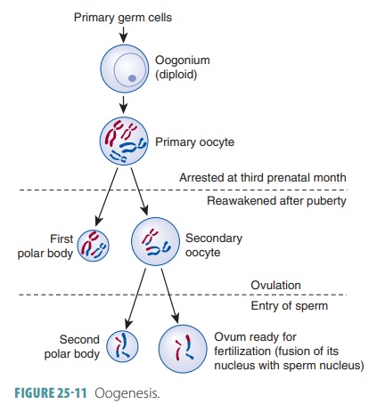
The first polar body may continually develop and undergo meiosis II, with two smaller polar bodies being produced. The secondary oocyte stops func-tioning in metaphase II, and this is the cell that is ovulated. When no sperm penetrates an ovulated sec-ondary oocyte, it deteriorates. However, if penetration by a sperm occurs, the oocyte completes meiosis II quickly. This produces a tinysecond polar body and a large fertilized egg cell called a zygote. The joining of the egg and sperm nuclei constitutes fertilization.
The polar bodies soon degenerate. They allow for production of egg cells with massive amounts of cyto-plasm and abundant organelles that carry the zygote through its first cell divisions, still with the right num-ber of chromosomes.
Ovarian Cycle
The ovarian cycle is the monthly series of events linked to maturation of an egg. It has two consecutive phases: the follicular phase and the luteal phase. In the follic-ular phase, the dominant follicle is selected and starts to secrete significant amounts of estrogens. This lasts from day one to 14, typically followed by ovulation.
Interestingly, only 10% to 15% of women actu-ally have 28-day cycles. The ovarian cycle may range between 21 and 40 days in actuality. When this is the case, there are variations in the length of the follicu-lar phase and the timing of actual ovulation. However, the luteal phase always begins on the 14th day after ovulation and lasts to the cycle’s end.
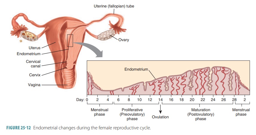
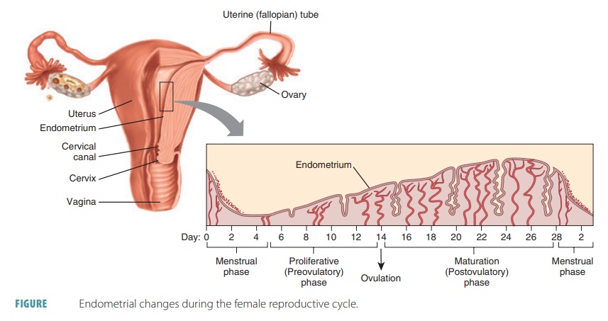
In younger females, estrogens stimulate devel-opment of the secondary sex characteristics. They maintain and develop these characteristics as time passes. Increased estrogens during the first week of a reproductive cycle thicken the glandular endo-metrium of the uterine lining. This is known as the proliferative phase. FIGURE 25-12 shows the various phases of the female reproductive cycle.
Follicular Phase
The first part of the follicular phase is known as the pre-antral phase, which is not dependent on gonadotropin. This is when cytokines, growth factors, and other intra-follicular paracrines control development of oocytes and follicles. The second antral phase is controlled by FSH and LH. Activated follicles grow greatly, and the primary oocyte in the dominant follicle restarts meiosis I.
In the follicular phase, the developing follicle matures, and by approximately day 14 of the cycle, it appears on the surface of the ovary as a blister-like bulge. Follicular cells inside the follicle loosen and fol-licular fluid accumulates. The maturing follicle secretes estrogens that inhibit the anterior pituitary from releasing LH but allow it to be stored. Anterior pituitary cells become more sensitive to GnRH secreted from the hypothalamus in rhythmic pulses. The stored LH is released, weakening and rupturing the bulging follicu-lar wall. This sends the secondary oocyte and fluid from the ovary in the process of ovulation. The space con-taining the follicular fluid fills with blood, which clots.
Near the end of follicle maturation, the primary oocyte finishes meiosis I. A secondary oocyte and first polar body are formed, readying the cycle for ovulation. The granulosa cells signal the oocyte to stop the comple-tion of meiosis. Follicle growth from the primordial stage to this point is believed to take about one year. Therefore, each follicle that ovulates was actually beginning to grow between 10 and 12 ovarian cycles previously.
Ovulation
The primary oocyte undergoes oogenesis, developing a secondary oocyte and first polar body. This is known as ovulation. This process releases these developed structures along with one or two layers of follicular cells from the mature follicle. Anterior pituitary gland hormones trigger ovulation, swelling the mature folli-cle while weakening its wall. The wall ruptures, allow-ing the fluid and secondary oocyte to be expelled into the peritoneal cavity while still surrounded by the corona radiata. There may be a slight pain in the lower abdomen at the moment this occurs. Although the cause is unknown, it may be due to extreme stretching of the ovarian wall and irritation of the peritoneum by blood or fluid from the ruptured follicle.
Every adult woman has several follicles that are continually at different stages of maturation. Because of this, one follicle is at the perfect stage of maturation when LH stimulates ovulation. Antral follicles sur-vive because of FSH, which helps to select the dom-inant follicle, although this actual process is not fully understood. It is believed to add the largest amount of gonadotropin receptors, attaining the most FSH sensitivity at the fastest rate. Other follicles undergo apoptosis and are reabsorbed by the body.
Luteal Phase
The follicular cells enlarge to form a temporary corpus luteum after the ruptured follicle or corpus hemorrhagicum is absorbed. Thisluteal phase is when the corpus luteum is active. Corpus luteum cells secrete large amounts of progesterone and estrogens during the last half of the cycle, and blood proges-terone concentration increases sharply. Progesterone causes the endometrium to become more vascular and glandular while stimulating uterine gland secre-tion of more lipids and glycogen. This is known as the secretory phase. Endometrial tissues fill with fluids that are made up of nutrients and electrolytes, which support embryo development.
LH and FSH release is then inhibited, and no other follicles develop when the corpus luteum is active. If no egg cell is fertilized, on the 24th day of the cycle the corpus luteum begins to degenerate, to be replaced by connective tissue. The leftover remnant is called a cor-pus albicans. Then, estrogens and progesterone decline in level, and the endometrium constricts its blood vessels. The uterine lining starts to disintegrate and slough off. Damaged capillaries create a flow of blood and cellular debris, which passes through the vagina. This is called the menstrual flow. It usually begins approximately on the 28th day of the cycle, continuing for three to five days while estrogen concentrations are low. TABLE 25-4 summarizes the female reproduc-tive cycle. However, if the oocyte is fertilized, resulting in pregnancy, the corpus luteum continues to develop until the placenta assumes its hormone production duties. This occurs in approximately three months.
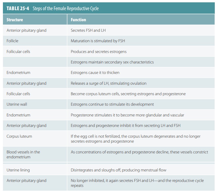
1. Distinguish the function of estrogens and progesterone in the female reproductive system.
2. What is menarche?
3. What is a corpus luteum?
Hormonal Regulation of the Ovarian Cycle
The female reproductive system is controlled by hor-mones, involving interplay between the pituitary gland and gonadal secretions. Female hormonal regu-lation is much more complicated than male hormonal regulation because it coordinates both the ovarian and uterine cycles.
The maturation of female sex cells, develop-ment and maintenance of secondary sex character-istics, and changes during the monthly reproductive cycle is controlled by the hypothalamus, anterior pituitary gland, and ovaries. Until about age 10, the female body is reproductively immature. When the hypothalamus begins to secrete more GnRH, the anterior pituitary releases FSH and LH, controlling female sex cell maturation and producing female sex hormones. The ovaries, adrenal cortices, and placenta secrete sex hormones during pregnancy that include estrogens and progesterone. The most abundant of the estrogens is estradiol, fol-lowed by estrone and estriol. Also, onset of puberty is linked to amounts of adipose tissue. Leptin is the hormone that informs the hypothalamus about these amounts. Puberty is delayed if blood levels of lipids and leptin are low.
In childhood, the ovaries are growing with con-tinuous secretion of only a small amount of estrogens. These keep the hypothalamus from releasing GnRH. With normal leptin levels, the hypothalamus becomes less sensitive to estrogen as puberty approaches. It starts to release GnRH rhythmically, with this hor-mone stimulating the anterior pituitary to release FSH and LH. As a result, the ovaries begin to secrete estrogens and other hormones in higher quanti-ties. Gonadotropin levels increase continuously for approximately four years. Females at this stage are still not ovulating and pregnancy is not possible. In non-pregnant females, the ovaries are the main source of estrogens. Hormonal interactions stabilize eventually and the adult ovarian cycle begins.
Estrogens and related hormones stimulate enlargement of accessory sex organs and develop and maintain the female secondary sex characteristics:
■■ Development of breasts and the mammary gland ductile systems
■■ Increasing adipose tissue deposition in the subcutaneous layer, breasts, thighs, and buttocks
■■ Increasing skin vascularization
The ovaries are also the main source of progesterone in nonpregnant females. Progesterone promotes uterine changes during the monthly cycle, affects the mammary glands, and helps regulate gonadotropin secretion. Con-centrations of androgen in females at puberty produce different changes, including increased hair growth in the pubic region and armpits. The female skeleton responds to low androgen concentration by narrowing the shoul-ders and widening the hips.
Uterine Cycle
Controlled by estrogen, the uterine glands, blood vessels, and epithelium change with the phases of the menstrual cycle, which is also called the uterine cycle. It is coordinated with the ovarian cycle. The changes in this cycle can be divided up into a menstrual phase, proliferative or preovulatory phase, and a secretory, postovulatory phase (FIGURE 25-13).
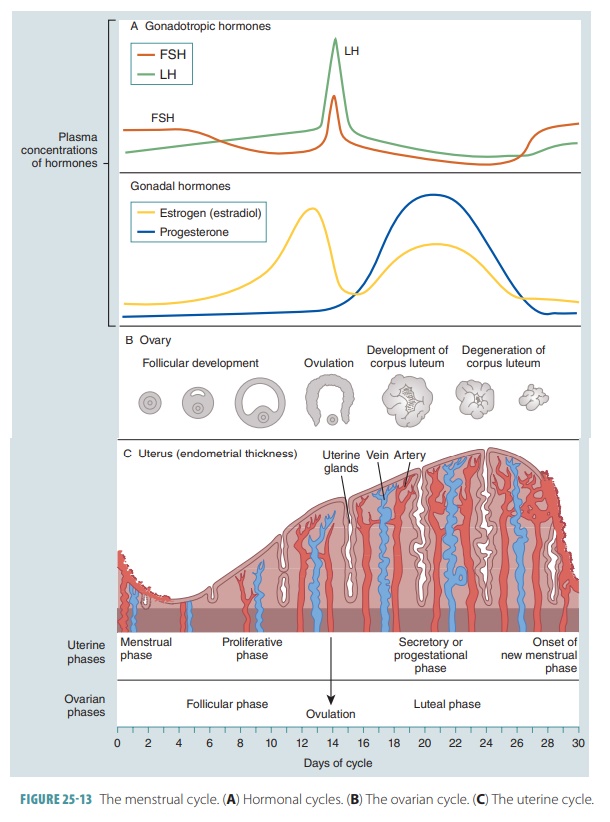
The menstrual phase occurs during days one to five. The uterus sheds all except the deepest part of its endometrium. Ovarian hormones are at their low-est normal levels, but gonadotropins are increasing. The functional layer of the endometrium is thick and depends on hormones. It detaches from the uterine wall, resulting in bleeding for three to five days. Blood and detached tissue flow out through the vagina. By day five, more estrogen is being produced by the ovarian follicles as they grow.
The proliferative phase occurs during days six to 14. The endometrium is rebuilt, influenced by the rise of estrogens in the blood. Its basal layer gener-ates a new functional layer, which thickens. Its glands increase in size and the spiral arteries become more numerous. As a result, the endometrium returns to its earlier state: It is soft, smooth, thick, and well supplied with blood vessels. Estrogens cause the endometrial cells to synthesize progesterone receptors. This makes them ready for interaction with progesterone.
Cervical mucus is usually sticky and thick. However, as estrogen levels increase, it thins to form channels allowing sperm to pass into the uterus. Ovu-lation takes only five minutes, occurring on or about day 14 in the ovary as a response to a sudden release of LH by the anterior pituitary. The ruptured follicle is converted by LH to a corpus luteum.
The secretory phase occurs during days 15–28. This phase is not as variable as the others. The endo-metrium is readied for an embryo to be implanted. The corpus luteum increases progesterone levels, which affect the endometrium by causing the spiral arteries to become more extensive and by converting the functional layer into a secretory mucosa. In an effort to sustain an embryo, the endometrial glands become larger and coiled. They secrete nutrients into the uterine cavity.
A cervical plug is formed as progesterone levels rise and make the cervical mucus viscous once more. The plug helps to block entry of pathogens, other for-eign materials, and sperm. Progesterone also helps to ready the uterus for the task of supporting an embryo. Increasing levels of progesterone and estrogen inhibit LH release from the anterior pituitary.
When no fertilization occurs, the corpus luteum degenerates, LH blood levels are reduced, progester-one levels are decreased, and the endometrium no longer has the hormones it needs to support a preg-nancy. Its spiral arteries become kinked and spasm. The ischemic endometrial cells die because of a lack of oxygen and nutrients. The glands regress, preparing menstruation to begin on day 28. The spiral arteries constrict for a final time, suddenly relaxing and open-ing wide. Blood flows into the weak capillary beds and they fragment. The functional layer is then sloughed off and the uterine cycle begins again with this new menstrual flow.
1. List the hormones most important for the regulation of the ovarian cycle.
2. Which hormone, if lower than normal, will delay the onset of puberty?
3. Explain the three phases of the uterine cycle.
Estrogen, Progesterone, and Female Reproductive Function
Estrogens are to females what testosterone is to males. Both these hormones generate reproductive function. Two things happen when estrogen lev-els rise during puberty in a female’s body: Oogen-esis is promoted with follicle growth in the ovaries and anabolic effects occur in the female reproduc-tive tract. The tract is readied for supporting a pregnancy, with enhanced motility occurring in the uterine tubes and uterus. There is thickening of the vaginal mucosa and maturation of the external genitalia. Because of estrogens, girls between the ages of 11 and 12 experience “growth spurts,” which are more dramatic than those seen in boys. However, this is a shorter term process for females, because increasing estrogen levels also cause a faster closure of the epiphy-ses of the long bones. Girls usually reach their full height between the ages of 13 and 15, whereas boys usually reach their full height between the ages of 15 and 19.
Secondary sex characteristics included by estro-gen include breast development; widening and lightening of the pelvis for future childbirth; and increased deposition of subcutaneous fat, mostly in the breasts and hips. The various types of estrogen also help to maintain low total blood cholesterol lev-els and high -density lipoprotein levels. They facili-tate calcium uptake, keeping the skeleton’s density intact. All these effects begin in puberty, yet are not true secondary sex characteristics.
Progesterone helps to establish and regulate the uterine cycle. It causes changes to occur in the cer-vical mucus. Mostly in pregnancy but in other times as well, progesterone inhibits uterine motility and assists estrogen in preparing the breasts for lactation. The term progesterone actually means “for gestation.” In the majority of pregnancies, it is the placenta and not the ovaries that supplies most progesterone.
Female Sexual Response
The erectile tissues of the clitoris and vaginal entrance respond to sexual stimulation. Parasympa-thetic nerve impulses release nitric oxide to dilate the erectile tissues, increase blood inflow, and swell the tissues. The nipples become erect and the vagina expands and elongates. If sexual stimulation is sufficiently intense, parasympathetic impulses cause the vestibular glands to secrete mucus into the ves-tibule, moistening and lubricating the surrounding tissues and lower vagina. This facilitates insertion of the penis.
Just as in males, touch and psychological stim-ulation increase sexual excitement along autonomic nerve pathways. The clitoris responds to local stim-ulation, culminating in an orgasm if stimulation is sufficient. Females do not ejaculate, but do experi-ence increased muscle tension, raised blood pres-sure and pulse rate, and uterine contractions. Just before orgasm, the outer one -third of the vagina is engorged with blood. This increases friction on the penis, with orgasm initiating reflexes directed by the sacral and lumbar spinal cord. The muscles of the perineum and walls of both the uterus and uterine tubes contract rhythmically. This helps transport sperm through the female reproductive tract toward the upper uterine tubes.
Females experience intense pleasure that is fol-lowed by relaxation, which is the same for males, but do not have a refractory period after orgasm. Therefore, females can experience multiple orgasms during one sexual experience. However, orgasm is not required for conception. It is now understood that female libido is not primarily influenced by the male sex hormone testosterone. It is instead influenced by dehydroepiandrosterone, which is an androgen produced by the adrenal cortex.
1. Which hormones generate reproductive function in females?
2. Explain why girls experience growth spurts that are more dramatic but shorter in overall duration than boys.
3. Describe the female sexual response and identify differences between it and the male sexual response.


