Biology Chapter 21
Neural Control and Coordination

- Topic 1 Nervous System : An Overview
- Main Properties of Neural Tissue
- Topic 2 Human Nervous System
- Reflex Action and Reflex Arc
- Topic 3 Sensory Reception and Processing
The human body has several organs. These organs cannot perform their functions independently. In order to maintain homeostasis for the normal physiology of the human body, functions of these organs/organ systems in our body must be coordinated, so that they can work in proper manner.
Topic 1 Nervous System : An Overview
Coordination is the process through which two or more organs interact and complement the functions of one another. On the other hand, integration is a process, which makes two or more organs to work as a functional unit in harmony.
For instance, when we do exercise, we observe significant increase in the rate of respiration, heart beat, blood flow, sweating, etc., to meet enhanced need of nutrients and energy for increased activities of lungs, heart, muscles and many other body organs, when we stop exercising, we witness that the increased activities of lungs, heart, nerves, kidneys, muscles, etc., gradually return to normal. Thus, during exercise, functions of various organs of the body are coordinated and integrated.
In higher animals (including human), two types of systems have been developed for the control, coordination and integration, i.e., nervous system and endocrine system. The nervous system provides an organised network of point to point connections for quick neural coordination. The endocrine system provides chemical integration through hormones.
Neural System
The neural system is the control system of the body which consists of highly specialized cells called neurons. The sensory neurons detect and receive information from different sense organs (receptors) in the form of stimuli and transmit the stimuli to the Central Neural System (CNS) through sensory nerve fibers. In CNS the processing of information is done and a conclusion is drawn.
The conclusion is sent to different organs (effectors) through motor nerves. These effectors then show the response accordingly.
The structure of a neuron is seen to consist of two separate functional regions, or compartments – the cell body together with the dendrites as one region, and the axonal region as the other.
The neural or nervous system is present in most of the multicellular animals. Its complexity increases from lower to higher animals.
Invertebrates have relatively simpler nervous system than the vertebrates.
Human Neural System
The whole nervous system of human being is derived from embryonic ectoderm.
The human neural system is divided into two parts
(i) the Central Neural System (CNS)
(ii) the Peripheral Neural System (PNS)
The CNS includes the brain and the spinal cord and is the site of information processing and control.
The PNS comprises of all the nerves of the body associated with the CNS (brain and spinal cord).
The nerve fibres of the PNS are of two types
(a) Afferent Fibres They transmit impulses from tissues/organs to the CNS.
(b) Efferent Fibres They transmit regulatory impulses from the CNS to the concerned peripheral tissues/organs.
The PNS is divided into two divisions i.e., somatic neural system and autonomic neural system.
The somatic neural system relays impulses from the CNS to skeletal muscles while, the autonomic neural system transmits impulses from the CNS to the involuntary organs and smooth muscles of the body.
The autonomic neural system is further classified into sympathetic neural system and parasympathetic neural system.
Neuron (Structural and Functional Unit of Neural System)
Neurons are the longest cells in the body. Human neural system has about 100 billion neurons. Majority of the neurons occur in the brain. Fully formed neurons never divide and remain in interphase throughout life.
A neuron is a microscopic structure composed of three major parts
1. Cell Body (Cyton or Soma)
Like a typical cell it consists of cytoplasm, nucleus and cell membrane. The cytoplasm has typical cell organelles like mitochondria, Golgi apparatus, rough endoplasmic reticulum, ribosomes, lysosomes, certain granular bodies, neurofibrils, neurotubules and Nissl’s granules.

Presence of neurofibrils and Nissl’s granules is the characteristic to all neurons. Neurofibrils play a role in the transmission of impulses.
2. Dendrites (Dendrons)
Dendrites are usually shorter, tapering and much branched processes that project out of the cell body. They also contain Nissl’s granules and may be one to several in number.
They conduct nerve impulses towards the cell body and are called afferent processes (receiving processes).

3. Axon
Axon is a single, usually very long process of uniform thickness. The part of cyton from where the axon arises is called axon hillock (most sensitive part of neuron).

The axon contains neurofibrils and neurotubules but does not have Nissl’s granules, cell organelles and granular bodies. The axon ends (distal end) in a group of branches, the terminal arborization (axon terminals).
When terminal arborisations of the axon meet the dendrites of another neuron to form a synapse, each branch terminates as a bulb-like structure called synaptic knobs, which possess mitochondria and secretory vesicles (containing chemicals called neurotransmitters). The axons transmit nerve impulses away from the cell body to a synapse or to a neuromuscular junction.
There are two types of axon
a. Myelinated
In myelinated nerve fibres Schwann cells form myelin sheath around the axon. The gaps between two adjacent myelin sheaths are called nodes of Ranvier. Myelinated nerve fibres are found in cranial and spinal nerves.

b. Non-myelinated
In non-myelinated nerve fibres Schwann cell does not form myelin sheath around the axon and are without nodes of Ranvier. They are commonly found in autonomous and somatic neural systems.
Types of Neurons on the Basis of Structure
Based on the number of axon and dendrites, the neurons are divided into three types
(i) Multipolar neurons These neurons have several dendrites and an axon. They are found in cerebral cortex.
(ii) Bipolar neurons These neurons have one dendrite and one axon. They are present in the retina of eye.
(iii) Unipolar neurons These neurons have cell body with one axon only. These are found usually in the embryonic stage.
Main Properties of Neural Tissue
The neural tissue has two outstanding properties
(a) Excitability It is the ability of nerve cells to generate an electrical impulse in response to a stimulus by altering the normal potential difference across their plasma membrane.
(b) Conductivity It is the ability of nerve cells to rapidly transmit the electrical impulse as a wave from the site of its origin along their length in a particular direction.
Functions of Neural System
The nervous system serves the following important functions
(i) Control and coordination Nervous system controls and coordinates the working of all parts of the body so that it functions as an integrated unit. This is achieved by three overlapping processes, i.e., sensory input, integration and motor output.
(ii) Memory Nervous system stores the impressions of previous stimuli and retrieves (recalls) these impressions in future. These impressions are referred to as the experiences or memory.
(iii) Homeostasis Nervous system helps in the maintenance of the body’s internal environment, i.e., homeostasis.
Generation and Conduction of Nerve Impulse
Nerve impulse is a wave of bioelectric/electrochemical disturbance that passes along a neuron during conduction of an excitation.
Impulse conduction depends upon
(i) Permeability of axon membrane (axolemma).
(ii) Osmotic equilibrium (electrical equivalence) between the axoplasm and Extracellular Fluid (ECF) present outside the axon.
The generation of a nerve impulse is the temporary reversal of the resting potential in the neuron.
It occurs in following three steps
Polarisation (Resting Potential)
In a resting nerve fibre (a nerve fibre that is not conducting an impulse), the axoplasm (neuroplasm of axon) inside the axon contains high concentration of K+ and negatively charged proteins and low concentration of Na+.
(i) In contrast, the fluid outside axon contains a low concentration of K+ and a high concentration of Na+ and thus form a concentration gradient.
(ii) These ionic gradients across the resting membrane are maintained by the active transport of ions by the sodium-potassium pump, which transports 3Na+ out wards and 2K+ inwards (into the cell).
(iii) As a result, the outer surface of the axonal membrane possesses a positive charge, while its inner surface becomes negatively charged and therefore, is polarized.
(iv) The electrical potential difference across the resting plasma membrane is called as the resting potential. The state of the resting membrane is called polarized state.
Depolarization (Action Potential)
When a stimulus of adequate strength (threshold stimulus) is applied to a polarized membrane, the permeability of the membrane to Na+ ions is greatly increased at the point of stimulation (site A).
(i) This leads to a rapid influx of Na+ followed by the reversal of the polarity at that site, i.e., the outer surface of the membrane becomes negatively charged and the inner side becomes positively charged. The polarity of the membrane at the site A is thus, reversed and said to be depolarized.
(ii) The electrical potential difference across the plasma membrane at the site A is called the action potential, another name of nerve impulse.
(iii) At adjacent sites, e.g., site B, the membrane (axon) has positive charge (still polarized) on the outer surface and a negative charge on its inner surface.
(iv) The stimulated negatively charged point on the outside of the membrane sends out an electrical current to the positive point next to it. As a result, a current flows on the outer surface from site B to site A, while on the inner surface current flows from site A to site B.
This process (reversal) repeats itself over and over again and a nerve impulse is conducted through the length of the neuron.
Re-polarization
(i) The rise in the stimulus-induced permeability to Na+ is extremely short-lived. It is quickly followed by a rise in permeability to K+.
(ii) Within a fraction of a second, Na+ influx stops and K+ outflow begins until the original resting state of ionic concentration is achieved. Thus, resting potential is restored at the site of excitation, which is called repolarization of the membrane. This makes the fibre once more responsive to further stimulation.
(iii) In fact until repolarization occurs neuron cannot conduct another impulse. The time taken for this restoration is called refractory period.
Note:
- When an impulse travels along a myelinated neuron, depolarization occurs only at the nodes of Ranvier. It leaps over the myelin sheath from one node to the next. This process, is called saltatory conduction.
- This process accounts for the greater speed of an impulse travelling along a myelinated neuron than along a non-myelinated one. It is up to 50 times faster than the non-myelinated nerve fibre.
- A nerve impulses is transmitted from one neuron to another through junctions called synapses. It is formed by the membranes of a pre-synaptic neuron and a post-synaptic neuron.
There are mainly two types of synapses
Electrical Synapses
(i) The membranes of pre and post-synaptic neurons are in very close proximity (i.e., in continuity). The continuity is provided by the gap junction (small protein tubular structures) between the two neurons.
(ii) In electrical synapse, there is minimal synaptic delay because of the direct flow of electrical current from one neuron into the other across these synapses.
Thus, impulse transmission across an electrical synapses is always faster than that across a chemical synapse. In such synapses, transmission of impulse is very similar to impulse conduction along a single axon.
(iii) Electrical synapses are rarely found in our system. It is found in cardiac muscle fibres, smooth muscle fibres of intestine and the epithelial cells of lens.
Chemical Synapses
The membranes of pre and post-synaptic neurons are separated by a fluid-filled space called synaptic cleft.
A brief description of the mechanism of synaptic transmission is given below
(i) When an impulse (action potential) arrives at a pre-synaptic knob, calcium ions from the synaptic cleft enter the cytoplasm of the pre-synaptic knob.
(ii) The calcium ions cause the movement of the synaptic vesicles to the surface of the knob.
The synaptic vesicles are fused with the pre-synaptic (plasma membrane and get ruptured (exocytosis) to discharge their contents (neurotransmitter) into the synaptic cleft.
(iii) The neurotransmitter of the synaptic cleft binds with specific protein receptor molecules, present on the post-synaptic membrane.
(iv) This binding action changes the membrane potential of the post-synaptic membrane, opening channels in the membrane and sodium ions to enter the cell. This causes the depolarization and generation of action potential in the post-synaptic membrane. Thus, the impulse is transferred to the next neuron.
(v) The new potential developed may be either excitatory or inhibitory.
Human Nervous System
The human neural system can be categorised to
(a) Central Nervous System (CNS)
(b) Peripheral Nervous System (PNS)
Central Nervous System (CNS)
It is the integrating and command centre of the nervous system which consists of the brain and spinal cord (as discussed earlier).
Brain
The brain is the central information processing organ of our body and acts as the ‘command and control system’.
It controls the following activities
(i) The voluntary movements and balance of the body.
(ii) Functioning of vital involuntary organs, e.g., Lungs, heart, kidneys, etc.
(iii) Thermoregulation, hunger and thirst.
(iv) Circardian (24 hrs) rhythms of our body.
(u) Activities of several endocrine glands and human behaviour.
(vi) It is also the site for processing of vision, hearing, speech, memory, intelligence, emotions and thoughts.
Location
The brain is the anterior most part of the central neural system, which is located in the cranium
(cranial cavity) of the skull.
Protective Coverings of the Brain
It is covered by three membranes or meninges (cranial meninges)
(i) The outermost membrane, the duramater is the tough fibrous membrane adhering close to the inner side of the skull.
(ii) The middle very thin layer called arachnoid membrane (arachnoid mater).
(iii) The innermost membrane, the piamater is thin, very
delicate, which is in contact with the brain tissue.
Note:
The human brain weights from 1200-1400 g. The human neural system has about 100 billion neurons, majority of them occur in the brain.
The human brain is divisible into three parts
(i) Forebrain (ii) Midbrain (iii) Hindbrain
i. The forebrain
It consists of Olfactory lobes The anterior part of the brain is formed by a pair of short club-shaped structures, the olfactory lobes. These are concerned with the sense of smell.
Cerebrum It is the largest and most complex of all the parts of the human brain. A deep cleft divides the cerebrum longitudinally into two halves, which are termed as the left and right cerebral hemispheres connected by a large bundle of myelinated fibres the corpus callosum.
The outer cover of cerebral hemisphere is called cerebral cortex. The cerebral cortex is referred to as the grey matter due to its greyish appearance (as neuron cell bodies are concentrated here).
The cerebral cortex is greatly folded. The upward folds, gyri, alternate with the downward grooves or sulci. Beneath the grey matter there are millions of medullated nerve fibers, which constitute the inner part of the cerebral hemisphere. The large concentration of medullated nerve fibres gives this tissue an opaque white appearance. Hence, it is called the white matter.
Lobes A very deep and a longitudinal fissure, separates the two cerebral hemispheres. Each cerebral hemisphere of the cerebrum is divided into four lobes, i.e., frontal, parietal, temporal and occipital lobes.
In each cerebral hemisphere, there are three types of junctional areas
Sensory areas receive impulses from the receptors and motor areas transmit impulses to the effectors.
Association areas are large regions that are neither clearly sensory nor motor in junction. They interpret the input, store the input and initiate a response in light of similar past experience. Thus, these areas are responsible for complex functions like memory, learning, reasoning and other intersensory associations.
Distction the posterioventral part of forebrain.
Its main parts are as follows
Epithalamus is a thin membrane of non-nervous tissue. It is the posterior segment of the diencephalon.
The cerebrum, wraps around a structure called thalamus, which is a major coordinating center for sensory and motor signalling.
The hypothalamus, that lies at the base of thalamus contains a number of centres, which control body temperature, urge for eating and drinking. It also contains several groups of neurosecretory cells, which secrete hormones called hypothalamic hormones.
The inner parts of cerebral hemispheres and a group of associated deep structures like amygdala, hippocampus, etc., form a complex structure (limbic lobe or limbic system) that are involved in the regulation of sexual behaviour,expression of emotional reactions, e.g„ excitement, pleasure,rage and fear and motivation,
ii. Midbrain
The midbrain is located between the thalamus hypothalamus of the forebrain and pons of the hindbrain. A canal called the cerebral aqueduct passes through, the midbrain.
The dorsal portion of the midbrain mainly consists of two pairs (i.e., four) of rounded swellings (lobes) called corpora quadrigemina.
iii. Hindbrain
The hindbrain consists of
(a) Pons consists of fibre tracts that interconnect different regions of the brain.
(b) Cerebellum is the second largest part of the human brain (means litde cerebrum). It has very
convoluted surface in order to provide the additional space for many more neurons.
(c) Medulla (oblongata) is connected to the spinal cord and contains centres, which control respiration, cardiovascular reflexes and gastric secretions.
Note:
- Midbrain and hindbrain form the brain stem. It is the posterior part of the brain that continues with the spinal cord.
- Out of the twelve pairs of cranial nerves (in higher vertebrates), ten pairs come from the brain stem.
Spinal Cord
(i) It forms the posterior part of the CNS, running mid-dorsally in the neural canal of the vertebral column. In an adult, the spinal cord is about 42-45 cm long. Its diameter varies at different levels.
(ii) The spinal cord is formed of two types of nervous tissue, i.e., grey matter and white matter.
(iii) The grey matter is surrounded by white matter, which consists of groups of myelinated axons.
(iv) The spinal nerve tracts are divisible into two, ascending (conducting sensory impulses towards brain) and descending (conducting motor impulses from brain).
(v) Spinal cord conducts impulses to and from the brain and controls most of the reflex activities and provides a means of communication between spinal nerves and the brain.
Reflex Action and Reflex Arc
The entire process of response to a peripheral nervous stimulation, that occurs involuntarily, i.e., without conscious effort or thought and requires the involvement of a part of the central nervous system is called a reflex action. The nervous pathway taken by nerve impulses in a reflex action is called reflex arc.
Types of Reflexes
Reflexes are categorised into two
(i) Unconditioned (inborn reflexes and transmitted through heredity) breast feeding and swallowing.
(ii) Conditioned (acquired after birth, i.e., adopted during the course of life time.) e.g., Withdrawl of a body part (like limb) which comes in contact with objects that are extremly hot, cold, pointed or animals that are scary or poisonous.
Mechanism of Reflex Action
(i) The reflex pathway comprises atleast, one afferent (receptor) neuron and one efferent (effector) neuron arranged in a series.
(ii) The afferent neuron receives signal from a sensory organ and transmits the impulse via a dorsal nerve root into the CNS (at the level of spinal cord).
(iii) The efferent neuron then carries signals from CNS to the effector. The stimulus and response in this way forms a reflex arc, e.g., Knee jerk reflex as shown above in the diagram.
Peripheral Nervous System (PNS)
The peripheral nervous system consists of
1. Somatic Neural System (SNS)
2. Autonomic Neural System (SNS)
1. Somatic Neural System
The somatic neural system contains nerves which relay impulses from CNS to skeletal muscles. These can be further categorised into cranial (from brain) and spinal nerves on the basis of their origin.
i.Cranial Nerves
These nerves emerge specifically from the forebrain and brain stem.
Note:
- Trochlear is smallest and thinnest nerve and possess difficulty in surgical operations.
- Trigeminal is also called dentist nerve. It is the largest cranial nerve. At its origin it is associated with ‘Gasserian Ganglion’.
- Facial nerve is associated with geniculate ganglion at its origin.
Their functions in comparative manner in a nut shell are given below
ii. Spinal Nerves
All spinal nerves are mixed, having sensory and motor fibres in approximately equal numbers. In humans, 31 pairs of spinal nerves are present as Cervical (8 pairs), Thoracic (12 pairs), Lumber (5 pairs), Sacral (5 pairs), Coccygeal (1 pair).
Note:
There are 10 pairs of cranial nerves in fishes and amphibians and 12 pairs in rest of the higher chordates.
There are 10 pairs of spinal nerves found in fishes and amphibians and 31 pairs in humans.
Based on their functions, the nerve fibres of PNS are divided into two groups, i.e., afferent fibres and efferent fibres.
The afferent nerve fibres transmit sensory impulses from tissues/organs to the CNS and form the sensory or afferent pathway. The efferent nerve fibres transmit motor impulses from CNS to the concerned tissues/organs and form the motor or efferent pathways.
2. The Autonomic Neural System (ANS)
The autonomic neural system consists of the sympathetic and parasympathetic nervous system. The former is called thoraco-lumber outflow and the latter is called craniosacral outflow depending upon their origin.
Sensory Reception and Processing
The sensory organs (receptors) enable us to detect all types of changes in the environment and send appropriate signals to the CNS, where all the inputs are processed and analysed. Signals are then sent to different centres of the brain.
The most complex sensory receptors consist of numerous sense cells, sensory neurons and associated accessory structures. For example, eye (sensory organ for vision) and the ear (sensory organ for hearing).
Eye
The organ of sight are a pair of eyes in human.
Position
The eyes are situated in the deep protective bony cavities, called the orbits or eye sockets of the skull.
Parts of an Eye
The adult human eye ball is nearly spherical in structure. It consists of tissues present in three concentric layers
(i) Outermost fibrous layer composed of sclera and cornea.
(ii) Middle layer consists of choroid, ciliary body and iris.
(iii) Innermost layer consists df retina.
Outermost Layer
(i) Sclera is an opaque outermost covering, composed of dense connective tissue that maintains the shape of the eyeball and protects all the inner layers of the eye.
(ii) Cornea is a thin transparent, front part of sclera, which lacks blood vessels but is rich in nerve endings.
Middle Layer
(i) Choroid is a pigmented layer (bluish) present beneath the sclera. It contains numerous blood vessels and nourishes the retina. The choroid layer is thin over the posterior two-thirds of the eye ball, but it becomes, thick in the anterior part to form the ciliary body.
(ii) The eye ball contains a transparent crystalline structure called lens. Ciliary body holds the lens in position, stretching and relaxation of ciliary body changes the focal length of the lens for accomodation.
(iii) Iris forms a pigmented circle of muscular diaphragm attached to the ciliary body in front of the lens. Its pigment gives eye its colour.
The movement of muscle fibres of iris controls the size (diameter) of pupil.
(iv) Pupil is the aperture surrounded by the iris. It contains two types of smooth muscles, circular muscles (sphincters) and radial muscles (dilators) of ectodermal origin.
(v) Sympathetic stimulation causes the radial muscles to contract and the pupil to dilate or get larger. Parasympathetic stimulation causes the circular muscles to contract and the pupil to constrict.
Inner Layer
The inner layer is the retina and it contains three layers of cells from inside to outside, i.e., ganglion cells, bipolar cells and photoreceptor cells.
The photoreceptors or visual cells are of two types, i.e., rods (rod cells) and cones (cone cells). Both of these cells contain light sensitive proteins called the photopigments.
The twilight (scotopic) vision is the function of the rods. These cells contain a purplish-red protein called the rhodopsin (visual purple), which contains a derivative of vitamin-A.
The daylight (photopic) vision and colour vision are functions of cones. There are three types of cones, which possesses characteristic photopigments that respond to red, green and blue lights.
The sensation of different colours are produced by various combinations of these cones and their photopigments. In case of equal stimulation of these cones, a sensation of white light is produced.
Optic Nerves
The optic nerves are connected with the brain. These nerves leave the eye and the retinal blood vessels enter it at a point medial to and slightly above the posterior pole of the eye-ball. Photoreceptor cells (rods and cones) are not present in that region and hence, it is called blind spot, as no image is formed at this spot.
Macula Lutea and Fovea Centralis
At the posterior pole of the eye lateral to the blind spot, there is a small oval, yellowish area of the retina called the macula lutea or yellow spot, which has at its middle a shallow depression, the fovea centralis (fovea).
The fovea is a thinned out portion of the retina where only the cones are densly packed. It is the point where the visual acuity (resolution) is the greatest.
Contents of the Eye
(i) Aqueous Humour The space between the cornea and lens is called the aqueous chamber, which contains a thin watery fluid called aqueous humour.
(ii) Vitreous Humour The space between the lens and retina is called the vitreous chamber, which is filled with a transparent get called the vitreous humour.
Mechanism of Vision
In human eyes, the vision is called binocular vision (i.e., both the eyes can be focused on a common object).
(i) Retina receives light rays (in visible wavelength) through the cornea and lens generate impulses in rods and cones.
(ii) The photosensitive compounds (photopigments) in the human eye are composed of opsin (a protein) and retinal (an aldehyde of vitamin-A).
(iii) The received light induces dissociation of the retinal from opsin resulting in changes in the structures of the opsin. This causes the changes in the permeability of membrane.
As a result, the potential differences are generated in the photoreceptor cells. This produces a signal that generates action potential in the ganglion cells through the bipolar cells.
(iv) These impulses (action potentials) are transmitted by the optic nerves to the visual cortex of the brain.
(v) In brain, neural impulses are analysed and the image formed on the retina is recognised (based on earlier
memory and experience).
Common Diseases
(i) Cataract This is a eye disease generally occur in older people (above 60 years). Lens becomes opaque due to disease or ageing. It leads to blindness. It can be corrected by wearing suitable glasses or by replacing the defective lens with a normal lens from a donor.
(ii) Myopia (near or short sightedness) It occurs due to convexity of lens or longer eye ball, which results in an image of distant objects being formed in front of the retina, and can be corrected by wearing spectables or concave lenses.
(iii) Hypermetropia (far or long sightedness.) The image of nearer object becomes blurred. It is due to image being formed beyond the retina due to eye ball being short or lens being flattened. It can be corrected by wearing convex or convergent lenses.
(iv) Presbiopia It generally occurs after 40 years. The loss of elasticity in the eye lens occurs so that near objects (written or printed words) are not correcdy visible. It can be correct’d by convex/bifocal lenses.
Ear
Ears are a pair of statiocoustic organs meant for both sensory functions, i.e., hearing and maintenance of body balance.
Position
The ears are located on the sides of the head.
In most mammals, the ear is a flap of tissue also called pinna. It is a part of auditory system.
The mammalian ear can be anatomically divided into three major sections
1. External Ear
The external ear consists of pinna and the auditory canal (external auditory meatus), which collect sound waves and channel them to tympanic membrane (ear drum) separating the outer ear from the middle ear.
The auditory canal leads inwards and extends upto the tympanic membrane (the ear drum).
There are very fine hairs and wax-secreting sebaceous glands in the skin of the pinna and the meatus. The tympanic membrane is composed of connective tissues covered with skin outside and with mucus membrane inside.
2. Middle Ear
The middle ear contains three ossicles called malleus (hammer), incus (anvil) and stapes (stirr-up), which are attached to one another in a chain-like fashion.
The malleus is attached to the tympanic membrane and the stapes is attached to the oval window (a membrane beneath the stapes) of cochlea.
These ossicles increase the efficiency of transmission of sound waves to the inner ear.
The middle ear also opens into the Eustachian tube, which connects with the pharynx and maintains the pressure on either sides of the ear drum. It also enables you to ‘pop’ your ears when you change altitude.
3. Inner Ear
The inner ear consist of a labyrinth of fluid-filled chambers within the temporal bone of the skull. The labyrinth consists of two parts the bony and membranous labyrinths. The bony labyrinth is a series of channels. Inside the channels, membranous labyrinth lies, which is surrounded by a fluid called perilymph.
The membranous labyrinth is filled with a fluid called endolymph. The coiled portion of the labyrinth is called cochlea.
The membranes constituting cochlea (the Reissner’s and basilar), divide the bony labyrinth into two large canals, i.e., an upper vestibular canal (scala vestibuli) and a lower tympanic canal (scala tympani).
These (both) canals are separated by a small cochlear duct called scala media. The vestibular and tympanic canals contain and the cochlear duct is filled with endolymph.
At the base of the cochlea, the scala vestibuli ends at the oval window while, the scala tympani terminates at the round window, which opens to the middle ear.
Organ of Corti
The floor of the cochlear duct, the basilar membrane bears the organ of Corti. It contains the mechanoreceptors of the ear. The hair cells are present in rows on the internal side of the organ of Corti, that act as auditory receptors. The basal end of the hair cell is in close contact with the afferent nerve fibres.
A large number of processes called stereo cilia are projected from the apical part of each hair cell. Above the rows of hair cells is a thin elastic membrane called tectorial membrane.
Vestibular Apparatus
(i) The inner ear also contains a complex system called vestibular apparatus (located above the cochlea). It is composed of three semicircular canals and the otolith organ consisting of the saccule and utricle.
(ii) Each semicircular canal lies in a different plane at right angles to each other. The membranous canals are suspended in the perilymph of the bony canals. The base of canals is swollen and is called ampulla, which contains a projecting ridge called crista ampullaris, which has hair cells.
(iii) The saccule and utricle contain a projecting ridge called macula. The crista and macula are the specific receptors of the vestibular apparatus responsible for the maintenance of balance of the body and posture.
Mechanisms of Hearing
(i) Sound waves from the environment are received by the external ear and it directs them to the ear drum.
(ii) The ear drum vibrates due to sound waves and the vibrations are send to oval window through the ear ossicles (malleus, incus and stapes).
(iii) The vibrations are passed through the oval window on to the fluid of the cochlea, where they generate waves in the lymph.
(iv) The waves in the lymph induce a ripple in the basilar membrane.
(v) These movements of the basilar membrane bend the hair cells, pressing them against the tectorial membrane. Due to this, the nerve impulses are generated in the associated afferent neurons. These impulses are transmitted by the afferent fibres via auditory nerves to the auditory cortex of the brain, where the impulses are analysed and the sound is recognised.
Common Diseases
(i) Meniere’s Syndrome It is a hearing loss due to pathological distension of membranous labyrinth.
(ii) Eustachitis It occurs due to inflammation of Eustachian tube.
(iii) Tympanitis It is due to inflammation of ear drum.
(iv) Otalgia Pain occurs in ear.
(v) Otitis media Acute infection in middle ear.
SHORT NOTES:
Neural System
The neural system of all animals is composed of neurons, which can detect, receive and transmit different types of stimuli. In the lower invertebrates, the neural system is very simple, composed of a network of neurons. The vertebrates have a more developed neural system.
Human Nervous System
The human nervous system is divided into two parts :
- The Central Neural System (CNS): Site of information processing and control. It includes:
- Brain
- Spinal cord
- The Peripheral Neural System (PNS): Consists of all the nerves associated with CNS. The nerve fibers of PNS are of two types
- Afferent fibers (transmit impulses from tissues/organs to the CNS)
- Efferent fibers (transmit regulatory impulses from the CNS to the concerned peripheral tissues/organs)
The PNS is divided into:
- Somatic Nervous System relays (transmits impulses from the CNS to skeletal muscles)
- Autonomic Nervous System(transmits impulses from the CNS to the involuntary organs and smooth muscles) It is further divided into the sympathetic neural system and parasympathetic neural system.
Visceral Nervous System
Part of PNS consists of a whole complex of nerves, fibers, ganglia, and plexuses by which impulses travel from the CNS to the viscera and from the viscera to the CNS.
Neurons
A neuron is a specialized cell that transmits information throughout the nervous system through electrical and chemical signals.
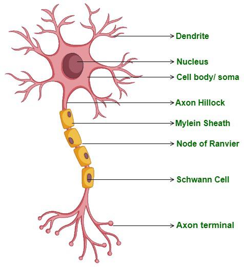
Diagram of Neuron
- A neuron is composed of three parts: cell body, dendrites, and axon.
- The cell body contains cytoplasm along with cell organelle and granules known as Nissl’s granules.
- Dendrites are small fibers that project out of the cell body. They also contain Nissl’s granules. Its function is to transmit impulses toward the cell body.
- Axon is a long fiber, branched at the distal end. The axons transmit nerve impulses away from the cell body to synapse or to a neuro-muscular junction.
- Neurotransmitters are present in the synoptic knob present at the end of the axon.
- Axons are divided into three types based on the number of axons and dendrites.
- Unipolar: cell body with one axon. Example: Embryonic stage.
- Bipolar: one axon and one dendrite. Example: Retina of the eye
- Multipolar: with one axon and two or more dendrites. Example: Cerebral cortex.
There are two types of axons, namely, myelinated and non-myelinated.

- The myelinated nerve fibers are enveloped with Schwann cells. It forms a myelin sheath around the axon. For example, Spinal and cranial nerves.
- Non-myelinated fiber is enclosed by a Schwann cell that does not form a myelin sheath around the axon. Example: Autonomous and somatic neural systems. Nodes of Ranvier are the gaps between two adjacent myelin sheaths.
Generation and Conduction of Nerve Impulse
First, let’s discuss why the membrane of the nerve is polarized. Neural membrane contains different types of ion channels, that are selectively permeable to different ions.
At the resting phase (when the neuron is not conducting any impulse), the axonal membrane is permeable to potassium ions and impermeable to sodium ions, and also to negatively charged proteins present in the axoplasm. Therefore, inside the axon, there is a high concentration of potassium ions and negatively charged proteins and a low concentration of sodium ions. Outside the axon, there is a low concentration of potassium ions and a high concentration of sodium ions. Thereby, creating a concentration gradient.
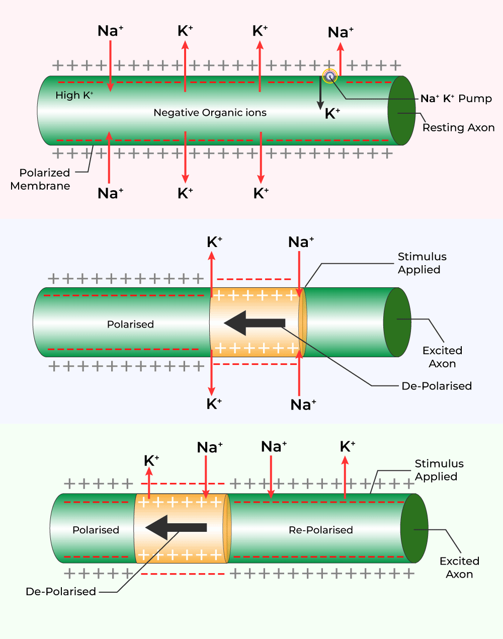
This ionic gradient is maintained by the sodium-potassium pump, which transports 3 Na+ outwards for 2 K+ into the cell. Hence, the axonal membrane’s outer surface possesses a positive charge, and the inner surface negative charge, therefore is polarized. The electrical potential difference across the resting plasma membrane is called the resting potential. Now let’s discuss the mechanism of conduction of impulse.
Mechanism of Generation and Conduction of Impulse
When the polarized membrane is stimulated, the membrane at site A becomes permeable to sodium ions, leading to a rapid influx of sodium ions, that results in the reversal of polarity at site A. Now, the outer surface of the membrane is negatively charged and the inner surface is positively charged. This reversal of polarity is known as depolarization. This difference in electrical potential across the membrane at site A is called a nerve impulse.

At site B, the outer surface of the membrane is positively charged, and the inner surface is negatively charged, therefore the current flows on the inner surface from site A to site B and on the outer surface from site B to site A, thus completing the circuit of current flow. The action potential is generated at site B, and the impulse generated at site A arrives at site B. The cycle is repeated along the length of the axon and an impulse is conducted.
Transmission of Nerve Impulses
Transmission of impulses from one neuron to another takes place through synapses. A synapse is formed by the membranes of a pre-synaptic neuron and a post-synaptic neuron, that may or may not be separated by a gap called synaptic cleft. Two types of synapses are:
Electrical synapses
The membrane of pre and post-synaptic neurons is very close. Current flows directly from one neuron to another in this synapse. This type of synapse is very rare in humans and is similar to the transmission in a single axon.

Chemical Synapses
The membranes of the pre and post-synaptic neurons are separated by a fluid-filled space called synaptic cleft. Neurotransmitters at the axonal ends are involved in the transmission of impulses at these synapses.
Neurotransmitter released at the synaptic cleft binds with the specific receptor, present on the post-synaptic membrane. New potential ( either excitatory or inhibitory) is generated at the postsynaptic neuron by opening ion channels.
Central Nervous System
The brain is present inside the skull and is covered by a membrane known as cranial meninges. Meninges is divided into three parts, the outer layer called the dura mater, the middle layer called the arachnoid, and the inner layer called pia mater. The brain performs various functions like balancing of body, thermoregulation, hearing, vision, voluntary movements, etc. The brain is divided into three parts, that is forebrain, midbrain, and hindbrain.
Also Read: Difference Between Brain and Spinal Cord Meninges
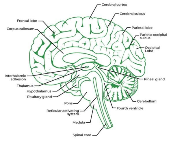
Forebrain
The forebrain consists of the cerebrum, thalamus, and hypothalamus.
- Cerebrum
- The cerebrum consists of left and right cerebral hemispheres connected by nerve fibers called the corpus callosum.
- The outer layer covering the cerebral hemispheres is called the Cerebral cortex or Grey matter.
- Within the cerebral cortex are motor areas, sensory areas, and large association areas.
- Beneath the cerebral cortex lies the white matter, consisting of fibers covered with a myelin sheath.
- Thalamus helps in the coordination of sensory and motor signals.
- Hypothalamus controls body temperature, urges for drinking and eating, and secret hypothalamic hormones.
Also Read: Difference Between Cerebellum And Cerebrum

Human Brain Diagram
Midbrain
Located between the thalamus/Hypothalamus of the forebrain and the pons of the hindbrain. The four lobes present at the dorsal portion of the midbrain are known as corpora quadrigemina.
Hindbrain
Consists of the pons, cerebellum, and medulla oblongata.
- Pons connects different regions of the brain through fiber tracts.
- Cerebellum provides space for more neurons through its convoluted surface.
- Medulla oblongata controls cardiovascular reflex, respiration, and gastric secretion.
Midbrain, pons, and medulla oblongata together form the brain stem. The brain stem connects the brain and spinal cord.
FAQs on Neural Control and Coordination
What is the Basic Unit of the Neural System?
The neuron is the basic unit of the neural system.
Give Parts of the Neuron.
The neuron consists of three parts as follows – Cell body consisting of cytoplasm and Nissl’s granules., Dendrites which are short fibers projecting from the cell body and Axon which is a long fiber, branched at the distal end.
Which Part of our Central Neural System Acts as a Master Clock?
The Hypothalamus of the central neural system acts as a master clock.
What is a Synapse?
A synapse is formed by the membrane of pre – synaptic neurons and post-synaptic neurons. These are of two types, Electrical synapses, and chemical synapses.
What is Neural Control and Coordination Class 11?
Neural control and coordination refer to the regulation and integration of bodily functions through the nervous system, involving the transmission of electrical signals between neurons to govern various physiological processes.
OTHER IMPORTANT QUESTIONS :
1. Briefly describe the structure of the following:
(a) Brain (b) Eye (c) Ear
Solution:
The structure is as follows:
(a) Structure of the brain

1. The brain is the central information processing organ of the body, acting as the ‘command and control system’. It is protected in the skull.
2. It is covered by three membranes known as cranial meninges – the outer layer is the dura mater which is a fibrous and tough membrane, the middle layer is the arachnoid, which is delicate and thin, the innermost layer is the pia mater which is an extension of the brain tissue. This layer is extremely vascular and supplied richly with blood
3. The three main regions of the brain are:
(i) Forebrain
(ii) Hindbrain
(iii) Midbrain
Forebrain – has three main parts – cerebrum, hypothalamus, thalamus
→Cerebrum forms the most important and major part of the entire brain. It is longitudinally segregated into halves by a deep cleft, each half is known as the cerebral hemisphere. Both these hemispheres are linked by the corpus callosum, which is a tract of nerve fibres. The cerebral hemispheres are internally hollow and the walls of the cerebrum have an inner medulla and an outer cortex.
The cerebral cortex consists of cell bodies of neurons which imparts a grey appearance; hence it is referred to as grey matter. The grey matter has many grooves (sulci) and folds (gyri). The higher the number of convolutions, the greater the intelligence.
The cerebral cortex consists of sensory areas, motor areas and association areas (neither motor nor sensory). These specific areas are responsible for complex functions, namely communication, memory and intersensory associations.
The cerebral medulla is made of axons of nerve fibres, which impart a white appearance. Hence it is referred to as white matter. There is a group of interrelated deep structures inside the cerebral hemispheres, namely the amygdala and hippocampus, which results in the formation of a complicated structure known as the limbic system or the limbic lobe.
Role – The cerebrum is the centre of memory, intelligence, consciousness, voluntary actions and willpower
→Thalamus
It is made up of grey matter and is located superior to the midbrain.
Role – it relays motor and sensory impulses to the cerebrum and also controls the manifestation of emotions, and comprehends heat, pain and cold.
→Hypothalamus
Located at the base of the thalamus, it consists of the optic chiasma. It is a point wherein the optic nerve fibres cross opposite sides. Behind this structure is the infundibulum, which is a greyish protuberance of the hypothalamus. It contains the pituitary gland.
Role – The hypothalamus has centres responsible for regulating the temperature of the body, homeostasis, and blood pressure, and the centre for controlling appetite (hunger, sleep, fatigue, thirst, pleasure, anger and penance). The neurosecretory cells of the hypothalamus produce releasing factors or several hormones that are crucial in regulating the activities of the pituitary hormones. Along with the limbic system, the hypothalamus also plays a part in regulating sexual behaviour.
Midbrain
It consists of the cerebral peduncles and the corpora quadrigemina
→Cerebral Peduncles
They are fibrous thick tracts which connect the cerebrum and the cerebellum.
Role – Relay the sensory and motor impulses between the hindbrain and the forebrain
→Corpora quadrigemina
The dorsal part of the brain has two pairs of solid lobes which are referred to as the corpora quadrigemina where one pair is referred to as the superior colliculi, and the other pair is referred to as the inferior colliculi
Role – Corpora quadrigemina controls the visual reflexes and the movement of the eye and head. They also regulate auditory reflexes and movement of the head to identify and detect the source of sound.
Hindbrain
It consists of the cerebellum, pons varolii and medulla oblongata
→Cerebellum
Present behind the top part of the brain stem. The outer cerebellar cortex consists of grey matter, and the inner cerebellar medulla consists of white matter. The cerebellum is connected with the medulla oblongata and the cerebrum through the fibre tracts of the white matter.
Role – It coordinates the balance of the body and muscular activity. The impulse to perform muscular activities is initiated in the cerebrum. It controls the voluntary movements originating in the cerebrum.
→Pons varolii
It is built of a thick bundle of white nerve fibres that are found above the medulla oblongata.
Role – Synchronizes between both the lobes of the cerebellum. It has the centre to control breathing which is referred to as the pneumotaxic centre.
→Medulla oblongata
It is conical in shape and is located at the skull’s base. It runs behind the brain as the spinal cord. Any injury to this site of the brain could be fatal.
Role – serves as a passage to conduct nerve impulses from the spinal cord to the brain. It controls all the activities of the internal organs, breathing and heartbeat.
(b) Structure of the Eye –

The human eye is embedded in a bony socket of the skull, and they are spherical. The walls of the eyeball consist of three layers, namely – the inner neurosensory coat, the middle vascular coat and the outer fibrous coat.
Outer fibrous coat – it is a thick and tough covering protecting the eyeball and helping to maintain its form. It has two regions – cornea and sclera
Sclera – consists of a dense white fibrous connective tissue, where only the white eye is visible, and the remaining major part is orbiting. The white of the eye is composed of collagen fibres.
Role – maintaining and protecting the shape of the eyeball.
Cornea – It is the non-vascular transparent part of the outer fibrous coat that is visible and is covered by a thin, transparent vascular layer of stratified epithelium known as the conjunctiva. It is in the continuation with the lining of the eyelids.
Role – The cornea refracts light which enters the eye and converges it into the lens.
Middle vascular coat – it consists of three regions, namely – choroid, ciliary body, and iris.
Choroid –
Highly vascular and made of loose fibrous connective tissue. It is in the continuation of the inner portion of the sclera. It finely layers over the posterior two-thirds of the eyeball and tends to turn thicker towards the front, imparting a bluish appearance. It consists of some pigmented cells.
Role – nourishes the retina supplying it with oxygen. The pigmented cells absorb excessive light to avoid reflection in the eyeball.
Ciliary body –
It is thick and forms the anterior part of the choroid. Comparatively, it is less pigmented and vascular and is composed of ciliary muscles and ciliary processes.
→Ciliary muscles – They are smooth muscles and are of two types – circular muscles and meridional muscles.
→Ciliary processes – the inner portion of the ciliary body has plenty of folds known as ciliary processes.
Role – secrete aqueous humour.
Iris – it is a fine, opaque and pigmented structure located at the junction of the sclera and cornea. The colour of the iris is imparted by the pigmented cells of the choroid. The colour varies between black, dark brown, blue or green. It consists of a pupil centrally as an aperture. The iris consists of two types of smooth muscles – circular muscles and radial muscles.
Role – iris controls the eye size and hence the amount of light that enters. When radial muscles contract, the pupil enlarges in dim light. When circular muscles contract, the pupil diminishes in bright light
Inner neurosensory coat –
The retina forms the innermost, neurosensory fine layer of the eyeball. The outer surface of the retina is in contact with the choroid, and the inner surface is in contact with the vitreous humourf.
The external surface has four layers:
Pigmented layer – it is made up of a single layer of cells containing dark-brown pigment.
The layer of photoreceptors – has two types of cells – rods and cones
Rods – it is rod-shaped and elongated, containing a purplish-red protein pigment known as rhodopsin or visual purple. It contains vitamin A derivatives. Rods do not respond to colours and are sensitive to dim light. They provide vision in the dark, hence known as twilight vision or scotopic vision
Cones – these are sensitive to colours and bright lights, providing daylight or photopic vision. The pigment that is found in the cone cells is known as iodopsin. Three kinds of cone cells respond to green, red and blue light. Other colours are detected by the simultaneous trigger of cone cells of more than one kind. A sensation of white light is generated when all three types of cells are simultaneously triggered.
Cone cells are insensitive to dim light hence in the dark, colour cannot be recognized.
A layer of bipolar neurons and a layer of ganglionic cells are the two layers. This layer contains the cell bodies of the ganglion cells, which forms the optic nerve.
Blind spot – The optic nerve exits the brain, retinal blood vessels enter the brain at a point where the photoreceptor cells are absent, this is the blind spot.
Macula lutea – It is the yellowish-pigmented spot that is present lateral to the blind spot. It is located exactly opposite the cornea’s centre. It has a pit located centrally known as the fovea, which lacks rods and blood vessels. It has only cone cells and is the area of most distinct vision.
Lens – It is elastic, transparent and biconvex in nature and is located just behind the iris. It is covered by a thin, elastic and transparent membrane known as the lens capsule. The lens is intact in its position due to the suspensory ligaments. These ligaments, along with the lens, segregate the eyeball into two chambers known as the aqueous chamber and the vitreous chamber.
Aqueous chamber – it is the space between the cornea and the lens containing a thin watery fluid known as aqueous humour.
Vitreous chamber – it is the space between the lens and the retina, which is filled with a transparent gel known as vitreous humour.
Ear


The human ear has two sensory functions – it enables hearing and maintains the balance of the body. It can be divided into three main sections – inner ear, outer ear, and middle ear.
→Inner ear – also known as the labyrinth, it is split into the membranous labyrinth and the bony labyrinth. The membranous labyrinth is filled with endolymph, while the bony labyrinth is filled with perilymph.
The membranous labyrinth is segregated into two portions – the vestibular apparatus and the cochlea. The vestibular apparatus consists of three semi-circular canals and an otolith. Each semi-circular canal lies in a different plane at right angles to each other. The membranous canals are suspended in the bony canals(perilymph). The base of the canals is swollen and is known as an ampulla containing crista ampullars – a projecting ridge which has hair cells.
The utricle and the saccule have a projecting ridge known as the macula. The macula and the crista are the particular receptors of the vestibular apparatus that have a role to play in maintaining posture and body balance.
Sacculus has a coiled and long outgrowth – cochlea, which is the chief hearing structure consisting of three membranes. A hearing organ, the organ of corti, is situated on the basilar membrane possessing hair cells.
→Outer ear – it has the pinna and the external auditory canal (meatus). The pinna gathers the vibrations in the air that generate sound. The external auditory canal extends up to the eardrum (tympanic membrane). It has very fine hair, wax-secreting glands in the skin of the meatus and the pinna. The tympanic membrane consists of connective tissues covered with a mucous membrane inside and with skin on the outside.
→Middle ear – it consists of three ossicles known as the malleus, stapes and incus that are linked to one another in a chain pattern. The malleus is linked to the tympanic membrane, and the stapes is linked to the oval window of the cochlea. The ear ossicles increase the efficiency of the transmission of sound waves to the inner ear. The middle ear cavity is connected to the pharynx through the Eustachian tube, which aids in equalizing the pressure on both sides of the eardrum.
2. Compare the following:
(a) Central neural system (CNS) and Peripheral neural system (PNS)
(b) Resting potential and action potential
(c) Choroid and retina
Solution:
The comparison is as shown below:
(a) Central neural system (CNS) and Peripheral neural system (PNS)
(b) Resting potential and action potential
(c) Choroid and retina
3. Explain the following processes:
(a) Polarisation of the membrane of a nerve fibre
(b) Depolarisation of the membrane of a nerve fibre
(c) Conduction of a nerve impulse along a nerve fibre
(d) Transmission of a nerve impulse across a chemical synapse
Solution:
(a) Polarisation of the membrane of a nerve fibre:

Impulse conduction through an axon
A nerve fibre is said to be in a polarized state when it is at a resting phase. In this polarized state, the membrane of the nerve fibres undergoes a resting potential. Listed below are the steps that occur during the process of polarization of the membrane of a nerve fibre:
1. Initially, when a depolarized region of a nerve fibre becomes polarized, the count of K+ ions outside the nerve fibres is more, while the axon membrane has an excessive number of Na+ ions.
2. When the membrane starts to turn into a polarized state, it turns more permeable to the K+ ions and impermeable to the negatively charged proteins and the Na+ ions
3. The 2 K+ ions are passed to the axon by a sodium-potassium pump through active transport, while the 3 Na+ ions are passed outside the axon
4. The outer side turns electropositive while the inner side of the membrane turns electronegative due to the movement of potassium and sodium ions which causes the nerve fibre to be polarized.
(b) Depolarisation of the membrane of a nerve fibre

1. A nerve fibre is said to be in a depolarized state when it is triggered
2. In this state, an action potential is experienced by the membrane of the nerve fibre
3. During the process of depolarization of the membrane of the nerve fibre, the following steps take place:
(i) Axon has more concentration of K+ ions in a polarized state, and outside the axon, the Na+ concentration is more.
(ii) The permeability of the membranes of Na+ and K+ ions is reversed when the nerve fibre is triggered by the stimulus.
(iii) The permeability for Na+ ions by the membrane increases
(iv) A rapid influx of Na+ ions into the axon is observed
(v) Hence, the inner side of the membrane turns positively charged while the outer side of the membrane turns negatively charged
(vi) This causes depolarization of the membrane of the nerve fibre, resulting in it experiencing an action potential
(c) Conduction of a nerve impulse along a nerve fibre
1. When a nerve impulse is conducted across the nerve fibre, it takes place in an organized manner
2. During the conduction of an impulse on the nerve fibre, a portion is always depolarized while the adjacent region is polarized. In order for the impulse to advance, repolarization of the depolarization area occurs while the polarized area depolarizes, which continues across the entire length of the nerve fibre helping in the impulse conduction.
3. It takes place in the following stages:
(i) Let A be a site on the depolarized region, where the inner surface of the membrane is positively charged while the outer surface of the membrane is negatively charged
(ii) At site B, the adjacent region is polarized, where the outer surface is positively charged while the inner surface of the membrane is negatively charged
(iii) Therefore, the flow of current at site A is on the inner surface of the membrane from A to B, and at site B, the flow of current is on the outer surface from B to A. This completes the circuit of current flow.
(iv) This causes the site B to depolarize so as to conduct the impulse at site B
(v) Site A gets repolarized as soon as the impulse is conducted to site B
(vi) Assume a site C adjacent to site B, which will be polarized when the site B is in a depolarized state.

(d) Transmission of a nerve impulse across a chemical synapse

1. The membranes of the pre-synaptic neuron and the post-synaptic neuron form a synapse
2. A synaptic cleft is a synapse which may or may not be segregated by a gap
3. The pre-synaptic neuron and the post-synaptic neuron at a chemical synapse are separated by the synaptic cleft
4. The calcium ions present at the synaptic cleft enters the synaptic knobs located at the axon terminal of the pre-synaptic neuron when an impulse reaches the axon terminal
5. The synaptic knobs have the synaptic vesicles of the pre-synaptic neuron which advance towards the plasma membrane to fuse with it
6. In the synaptic cleft, the vesicles release the neurotransmitter acetylcholine
7. The acetylcholine molecules tend to bind to the protein receptors located on the plasma membrane of the post-synaptic neurons
8. The binding opens up channels for sodium ions to enter the post-synaptic neuron. Simultaneously, the potassium ions exit the post-synaptic membrane
9. This causes an action potential in the post-synaptic neuron membrane. Thus, the impulse is conveyed to the post-synaptic neuron.
4. Draw labelled diagrams of the following:
(a) Neuron (b) Brain (c) Eye (d) Ear
Solution:
The diagrams are as follows:
(a) Neuron

(b) Brain

(c) Eye

(d)Ear

5. Write short notes on the following:
(a) Neural coordination
(b) Forebrain
(c) Midbrain
(d) Hindbrain
(e) Retina
(f) Ear ossicles
(g) Cochlea
(h) Organ of Corti
(i) Synapse
Solution:
(a) Neural coordination –It is a phenomenon through which two or more organs interact and complement functionalities of each other through the neural system of the body. The various physiological processes that take place in the body are interlinked with each other. The neural and the endocrine system jointly are responsible for coordinating and integrating all the actions and activities of the organs such that they function in a synchronized manner. This neural system renders a systematic and organized network for a point-to-point connection for prompt coordination. The endocrine system renders chemical integration through the hormones.
(b) Forebrain –
Forebrain – has three main parts – cerebrum, hypothalamus, thalamus
→Cerebrum forms the most important and major part of the entire brain. It is longitudinally segregated into halves by a deep cleft, each half is known as the cerebral hemisphere. Both these hemispheres are linked by the corpus callosum, which is a tract of nerve fibres. The cerebral hemispheres are internally hollow, and the walls of the cerebrum have an inner medulla and an outer cortex.
The cerebral cortex consists of cell bodies of neurons which imparts the grey appearance; hence it is referred to as grey matter. The grey matter has many grooves (sulci) and folds (gyri). Higher the number of convolutions, the greater the intelligence.
The cerebral cortex consists of sensory areas, motor areas and association areas (neither motor nor sensory). These specific areas are responsible for complex functions, namely communication, memory and intersensory associations.
The cerebral medulla is made of axons of nerve fibres, which impart a white appearance. Hence it is referred to as white matter. There are a group of interrelated deep structures inside the cerebral hemispheres, namely the amygdala and hippocampus which results in the formation of a complicated structure known as the limbic system or the limbic lobe.
Role – The cerebrum is the centre of memory, intelligence, consciousness, voluntary actions and willpower
→Thalamus
It is made up of grey matter and is located superior to the midbrain.
Role – it relays motor and sensory impulses to the cerebrum and also controls the manifestation of emotions, and comprehends heat, pain and cold.
→Hypothalamus
Located at the base of the thalamus, it consists of the optic chiasma. It is a point wherein the optic nerve fibres cross opposite sides. Behind this structure is the infundibulum, which is a greyish protuberance of the hypothalamus. It contains the pituitary gland.
Role – The hypothalamus has centres responsible for regulating the temperature of the body, homeostasis, and blood pressure, and the centre for controlling appetite (hunger, sleep, fatigue, thirst, pleasure, anger and penance). The neurosecretory cells of the hypothalamus produce releasing factors or several hormones that are crucial in regulating the activities of the pituitary hormones. Along with the limbic system, the hypothalamus also plays a part in regulating sexual behaviour.
(c) Midbrain
It consists of the cerebral peduncles and the corpora quadrigemina
→Cerebral Peduncles
They are fibrous thick tracts which connect the cerebrum and the cerebellum.
Role – Relay the sensory and motor impulses between the hindbrain and the forebrain
→Corpora quadrigemina
The dorsal part of the brain has two pairs of solid lobes which are referred to as the corpora quadrigemina where one pair is referred to as the superior colliculi and the other pair is referred to as the inferior colliculi
Role – Corpora quadrigemina controls the visual reflexes and the movement of the eye and head. They also regulate auditory reflexes and movement of the head to identify and detect the source of sound.
(d) Hindbrain
It consists of the cerebellum, pons varolii and medulla oblongata
→Cerebellum
Present behind the top part of the brain stem. The outer cerebellar cortex consists of grey matter and the inner cerebellar medulla consists of white matter. The cerebellum is connected with the medulla oblongata and the cerebrum through the fiber tracts of the white matter.
Role – It coordinates the balance of the body and muscular activity. The impulse of the performing muscular activity is initiated in the cerebrum. It controls the voluntary movements originating in the cerebrum.
→Pons varolii
It is built of a thick bundle of white nerve fibers that is found above the medulla oblongata.
Role – Synchronizes between both the lobes of the cerebellum. It has the center to control breathing which is referred to as the pneumotaxic center.
→Medulla oblongata
It is conical in shape and is located at the skull’s base. It runs behind the brain as the spinal cord. Any injury to this site of the brain could be fatal.
Role – serves as a passage to conduct nerve impulses from the spinal cord to the brain. It controls all the activities of the internal organs, breathing and heartbeat.
(e) Retina
It is the innermost layer containing layers of neural cells, namely – ganglion cells, bipolar cells and photoreceptor cells (mentioned in order from inside to the outside). The photoreceptor cells are of two types – cones and rods. Cones are responsible to impart daylight vision or colour vision whereas rods impart twilight vision. Image of an object is formed on the retina when light enters through the cornea, the lens.
(f) Ear ossicles
The middle ear possesses three ear ossicles known as malleus, incus and stapes that are interlinked to one another in a chain-like pattern. The malleus is in contact with the tympanic membrane, the incus with stapes and the stapes in turn with the oval window of the cochlea. The ear ossicles promote and cause an increase in the efficiency of sound wave transmission to the inner ear.
(g) Cochlea
It is the coiled portion of the labyrinth. The membranes constituting cochlea, the basilar and Reissner’s segregate the enveloping perilymph that is filled with the bony labyrinth into an upper scala vestibule and a lower scala tympani. The scala media (space within the cochlea) is filled with endolymph, and at the base of the cochlea, the scala vestibule terminates at the oval window, whereas the scala tympani ends at the round window that opens to the middle ear.
(h) Organ of Corti
Situated in the basilar membrane of the inner ear, the organ of corti is the organ of hearing. It contains hair cells that have auditory receptor cells, which are in turn found in rows on the internal side of the organ. Stereo cilia are the processes that are found on the apical ends of the hair cells whereas the basal sections of the hair cells consist of synaptic contacts with afferent nerve fibers. Just above the rows of these hair cells, a smooth gelatinous layer known as the tectorial membrane is found.
(i) Synapse –It is formed by the membranes of a pre-synaptic and a post-synaptic neuron, that may or may not be segregated by a gap known as the synaptic cleft. There are two types of synapses, namely chemical synapses and electrical synapses.
6. Give a brief account of:
(a) Mechanism of synaptic transmission
(b) Mechanism of vision
(c) Mechanism of hearing
Solution:
(a) Mechanism of synaptic transmission:
Synapses are the junctions where nerve impulses are transmitted from one neuron to another. These are formed by the membranes of a pre-synaptic and a post-synaptic neuron, which may or may not be segregated by a gap known as the synaptic cleft. There are two types of synapses, namely chemical synapses and electrical synapses.
The membranes of a pre-synaptic and a post-synaptic neuron at the electrical synapses are in close proximity so that electrical current can directly flow from one neuron to the other across these particular synapses. The transmission of an impulse across electrical synapses is similar to the conduction of an impulse along a single axon, where the transmission is always quicker than that across a chemical synapse, which is not commonly observed in the human body.
The membranes of a pre-synaptic and a post-synaptic neuron at the chemical synapses are segregated by a synaptic cleft (fluid-filled space). The chemicals known as neurotransmitters are involved in impulse transmission at these particular synapses.
(b) Mechanism of vision:
The passage of light rays is as follows – pupil, lens, aqueous humour, vitreous humour and finally, retina. This light causes the dissociation of the photo-pigment rhodopsin to retinal and opsin. The structure of opsin is subjected to changes due to the dissociation of opsin from the retinal which generates an action potential in the cones and rods of the retina. Furthermore, the action potential is transmitted to the ganglion cells via the bipolar neurons and ultimately transmitted to the visual cortex of the brain through the optic nerve. Analysis of impulses takes place in the visual cortex; responses are sent back in order to form images on the retina.
(c) Mechanism of hearing:
The pinna of the external ear collects sound waves, which pass through the external auditory meatus all the way to the eardrum. This causes the eardrum to vibrate. These vibrations are passed from the eardrum to the malleus, incuse and stapes of the middle ear which causes an increase in the frequency of the vibrations. The vibrations furthermore are passed to the cochlea of the inner ear through the oval window. These vibrations in the endolymph of the cochlea cause vibrations to be induced in the basilar membrane, which in turn causes the sensory hair of the organ of corti to vibrate.
The receptor hair cells force themselves against the tectorial membrane converting sound energy to a nerve impulse or action potential. This nerve impulse is transmitted to the auditory cortex of the brain, where the impulse is evaluated and analyzed, causing the sound to be recognized.
7. Answer briefly:
(a) How do you perceive the colour of an object?
(b) Which part of our body helps us in maintaining the body balance?
(c) How does the eye regulate the amount of light that falls on the retina?
Solution:
(a) Colour vision is imparted by the cone cells that are found in the retina of the eye. Cone cells are of three types which respond to red, green and blue light, respectively. At different wavelengths of light, different cone cells get triggered. Simultaneous stimulation of more than one kind of cone cells causes the other colours to be detected. A sensation of white light is observed when all three types of cone cells are triggered at the same time. This is how colour is perceived.
(b) The part of the body that helps in maintaining the body balance is the crista ampullaris which is located in the three semicircular canals, the macula sacculi found in the saccule of the inner ear and the macular utriculi found in the utricle.
(c) Pupil is an aperture located in the centre of the iris. Light enters the eye through this aperture.
The iris has two types of muscles, namely the radial smooth muscles and circular smooth muscles, that check the amount of light falling on the retina. The pupil diminishes in size when the smooth circular muscles contract in bright light. Therefore, less amount of light falls on the retina. The pupil widens when the light is dim due to the contraction of the radial smooth muscles such that enough light is incident on the retina.
8. Explain the following:
(a) Role of Na+ in the generation of action potential.
(b) Mechanism of generation of light-induced impulse in the retina.
(c) Mechanism through which a sound produces a nerve impulse in the inner ear.
Solution:
(a) Role of Na+ in the generation of action potential
When a nerve fibre is triggered, the sodium channels of the neurilemma are open and activated. From the outside, the sodium ions diffuse to the intracellular fluid due to the electrochemical gradient that is established. The membrane gets charged negatively from the outside as the potassium ions move out and gets positively charged from the inside. The immediate change that occurs in the membrane is known as action potential causing the membrane to get depolarized.
(b) Mechanism of generation of light-induced impulse in the retina
The human eye consists of photo pigments known as retinal and opsin. These are dissociated when light induces bringing about a change in the structure of opsin, causing an action potential to generate in the bipolar neurons. These action potentials or impulses are conveyed to the visual cortex of the brain by the optic nerves, where these impulses are read to analyze, and recognising an erect image.
(c) Mechanism through which a sound produces a nerve impulse in the inner ear
Vibrations are received through the membrane layering the fenestra ovalis by the perilymph of the internal ear. From here (the perilymph), vibrations are conveyed to the scala vestibule of the cochlea and, furthermore, to the scala media via the Reissner’s membrane, triggering the sensory hair of the organ of corti, which is the organ of hearing. These hair cells receive impulses to carry them to the brain through the auditory nerve, where the sense of hearing is felt.
9. Differentiate between:
(a) Myelinated and non-myelinated axons
(b) Dendrites and axons
(c) Rods and cones
(d) Thalamus and Hypothalamus
(e) Cerebrum and Cerebellum
Solution:
Listed below are the differences:
(a) Myelinated and non-myelinated axons
(b) Dendrites and axons
(c) Rods and cones
(d) Thalamus and Hypothalamus
(e) Cerebrum and cerebellum
10. Answer the following:
(a) Which part of the ear determines the pitch of a sound?
(b) Which part of the human brain is the most developed?
(c) Which part of our central neural system acts as a master clock?
Solution:
(a) The part of the ear that determines the pitch of a sound is the cochlea
(b) The part of the human brain that is the most developed is the cerebrum
(c) The part of the central neural system that acts as a master clock is the hypothalamus
11. The region of the vertebrate eye, where the optic nerve passes out of the retina, is called the
(a) fovea
(b) iris
(c) blind spot
(d) optic chaisma
Solution:
The region of the vertebrate eye where the optic nerve passes out of the retina is called the
(c) Blind spot
The optic nerves exit the eye, and the retinal blood vessels enter it at a point medial to and slightly above the posterior pole of the eyeball. Here, photoreceptor cells are absent and hence it is referred to as the blind spot.
12. Distinguish between:
(a) afferent neurons and efferent neurons
(b) impulse conduction in a myelinated nerve fibre and unmyelinated nerve fibre
(c) aqueous humor and vitreous humor
(d) blind spot and yellow spot
(e) cranial nerves and spinal nerves.
Solution:
The differences are as follows:
(a) Afferent neurons and efferent neurons
(b) Impulse conduction in a myelinated nerve fibre and unmyelinated nerve fibre
(c) Aqueous humor and vitreous humor
(d) Blind spot and yellow spot
(e) Cranial nerves and spinal nerves


No comments:
Post a Comment